Chronic upper back and rib pain in a healthy man: re-examining the cause
Cai Jun Jean Liang 1 4 , George Wen-Gin Tang 2 , Philip Herald 31 The University of New South Wales, Faculty of Medicine, NSW, Australia.
2 The University of New South Wales, Faculty of Medicine, School of Public Health and Community Medicine, NSW, Australia.
3 Waratah Private Hospital, Hurstville, NSW, Australia
4 Corresponding author. Email: jeaninlcj@gmail.com
Journal of Primary Health Care 13(2) 180-185 https://doi.org/10.1071/HC20027
Published: 28 May 2021
Journal Compilation © Royal New Zealand College of General Practitioners 2021 This is an open access article licensed under a Creative Commons Attribution-NonCommercial-NoDerivatives 4.0 International License
Abstract
INTRODUCTION: Back and rib pain is a common presentation in primary care practice. Although most cases are secondary to non-specific musculoskeletal pain, it is essential for clinicians to identify patients presenting with life-threatening pathologies.
AIM: This case report serves as a reminder to clinicians to reconsider their initial diagnosis when a patient’s pain fails to improve, while considering life-threatening pathologies.
CASE HISTORY: We describe a 44-year-old man from India who presents to his general practitioner with a 2-week history of rib and upper back pain. He was initially diagnosed with non-specific musculoskeletal pain. However, after representing twice 2 months later due to persistent pain and due to the uncertainty about his condition, he was investigated with different imaging modalities. It was discovered on bone scan that he had osteolytic lesions in the right 11th rib and T2 vertebrae. As the cause of his osteolytic lesions were unclear, he was referred to different specialists. Skeletal tuberculosis was suspected when one of his specialists discovered his recent visit to India, a tuberculosis-endemic country. This reminded the specialist of the possible risks of the patient’s background and its association with his symptoms. Bone biopsy of his lytic lesions revealed Mycobacterium tuberculosis, consistent with skeletal tuberculosis.
DISCUSSION: Revisiting the diagnosis of back and rib pain while considering other obscure and urgent pathologies is essential if a patient fails to improve clinically. Clinicians should focus on aspects of their clinical assessment to explore these pathologies, enabling earlier recognition of the disease.
Introduction
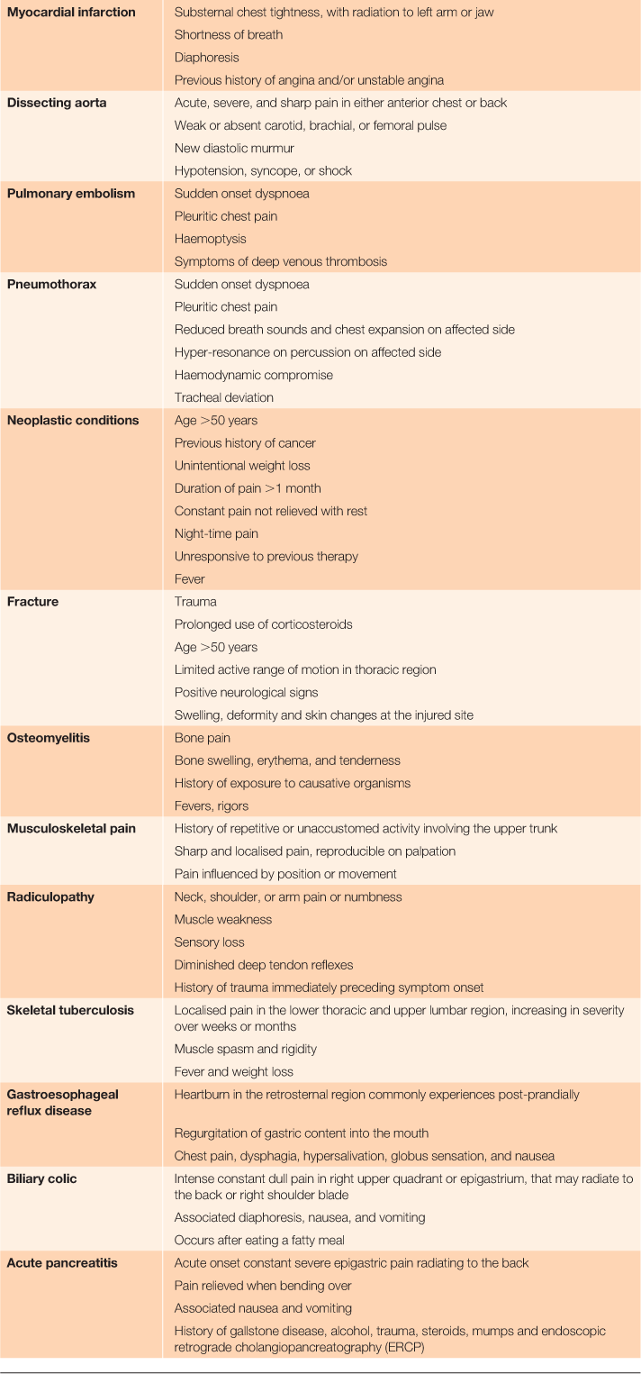
Back and rib pain is a common presentation that prompts patients to seek medical help. The differential diagnoses (Table 1) range from non-specific musculoskeletal pain occurring in most patients, to life-threatening pathologies in fewer patients.1 Although it is uncommon for patients to present with life-threatening pathologies, it is essential for clinicians to identify patients presenting with ‘red flags’.2–4 This case report describes the care of a patient who was initially treated for non-specific musculoskeletal pain, but his pain persisted for 10 weeks and was eventually attributed to a life-threatening pathology.

|
Case report
Presentation
A previously healthy 44-year-old Indian male presented to a Sydney General Practitioner (GP) in late 2017 with a 2-week history of a sudden onset, localised, sharp, constant lower right rib and left upper back pain. He also visited his regular GP at that time from his workplace in Bowral. The pain was worse at night and on movement and was not relieved by analgesia. He denied any preceding trauma to the area. There were no complaints of bladder and bowel dysfunction, paraesthesia, and weakness. He did not experience rash, fever, night sweats and weight loss. The patient denied any past medical and family history of cancers. In his initial history, there was no known recent travel. The patient was born in India and migrated to Sydney in 2009.
His vitals and gait were normal. There was no erythema, swelling and overlying skin changes on his chest. Localised tenderness was elicited on the right posterior 11th rib and left paraspinal muscles in the T4 region. Full range of motion was elicited in the cervical, thoracic, and lumbar spine. Heart sounds were dual and lung sounds were clear. His Bowral GP did an ECG a day earlier, which revealed normal sinus rhythm and ordered a chest X-ray and soft tissue ultrasound of his ribs and back, which were normal. As the initial impression was a non-specific musculoskeletal pain, the patient was treated conservatively.
Progress
The patient re-presented to his Bowral GP 1 month later with ongoing pain unrelieved by analgesia. A CT chest scan ordered by his Bowral GP revealed no abnormalities. He later re-presented to his Sydney GP a month later for his persistent pain. As his pain had been ongoing for 10 weeks, he was referred to a rehabilitation specialist for further assessment. Uncertain of the diagnosis, the specialist sent the patient for a bone scan.
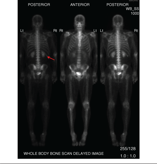
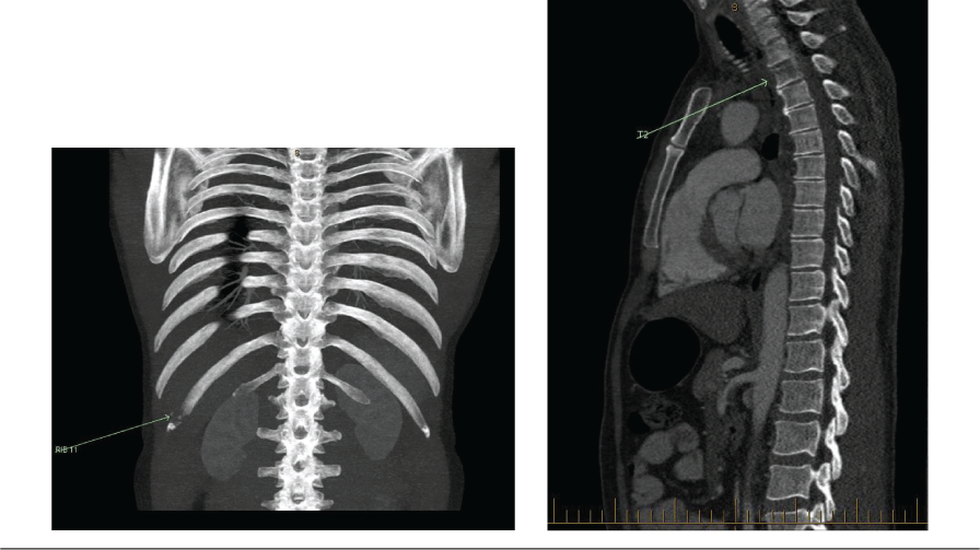
The patient’s bone scan (Figure 1) revealed an osteolytic process in the right 11th rib. As the initial CT scan had insufficient views visualising the patient’s ribs, a repeat CT chest, abdomen and pelvis scan (Figure 2) was performed; this demonstrated lytic lesions in T2 vertebral body and right posterolateral 11th rib, with no primary identified and prompted an urgent referral to an oncologist. The patient’s serological results for cancers were unremarkable. As the diagnosis was unclear, he was referred to a second oncologist. On detailed questioning by the second oncologist, the patient stated that 2 months before his presentation, he was in India, his home country, for a month. India is tuberculosis (TB)-endemic. Knowledge about his recent visit to India reminded the specialist of the risks of TB associated with the patient’s background. A provisional diagnosis of skeletal TB was made and the patient underwent a bone biopsy of his lytic lesions.

|

|
Results of his bone biopsy revealed Mycobacterium tuberculosis. The patient completed 9 months of anti-TB treatment and his pain improved significantly.
Discussion
Revisiting the initial diagnosis of non-specific musculoskeletal pain is essential when patients’ pain fails to improve. Clinicians should derive clues from the patient’s history to identify alternative pathologies causing pain. Despite being reviewed by two GPs and three specialists over a period of 2 months, only the last specialist made the association between the patient’s background and his risk of acquiring skeletal TB.
Most cases of extrapulmonary TB seen in Australia are from countries and regions outside Sydney.5,6 As Sydney is not an endemic region for TB, there was initially a low suspicion for skeletal TB with respect to the patient’s presentation, which accounted for the delayed diagnosis. However, this patient’s persistent pain should have prompted exploration of more obscure pathologies in relation to his background. As the patient was from India, skeletal TB should have been prioritised as a likely differential diagnosis.
Magnetic resonance imaging (MRI) is the best modality for characterisation of soft tissue extent of infection, including encroachment of adjacent organs and spinal cord. MRI is superior to imaging with CT.
Nuclear medicine bone scan is the most sensitive test for determining extent of active skeletal disease, as the entire skeleton is imaged; however, specificity is low, so areas of abnormal uptake on a bone scan need to be further assessed with other imaging modalities.
Computed tomography (CT) is the best modality for detecting and characterising infective bone lysis. CT can characterise extension of infection into the soft tissues including TB-associated calcification.
X-ray of the spine and ribs can demonstrate bone lysis from infection. Chest X-rays are good for looking for features of previous TB infection, but do not predict skeletal involvement.
The diagnosis of skeletal TB is challenging because there is no evidence of active chest disease in most patients, as in this case where a clear chest X-ray was reported. Other imaging modalities can be used to assist in the diagnosis of skeletal TB (Table 2).7,8 Suspicion of skeletal TB is usually derived from history focusing on the patient’s TB contacts.9 By focusing on the patient’s frequency of visiting India, skeletal TB would have been suspected much earlier.

|
Competing interests
The authors declare no competing interests.
Funding statement
This case report did not receive any specific funding.
Acknowledgements
We would like to thank the patient for sharing his medical details about his upper back and rib pain to allow other medical colleagues to learn from his case. Dr George Tang provided consistent guidance and patience to the first author to develop this case report.
References
[1] Maher C, Underwood M, Buchbinder R. Non-specific low back pain. Lancet. 2017; 389 736–47.| Non-specific low back pain.Crossref | GoogleScholarGoogle Scholar | 27745712PubMed |
[2] Yelland M, Cayley WE, Vach W. An algorithm for the diagnosis and management of chest pain in primary care. Med Clin North Am. 2010; 94 349–74.
| An algorithm for the diagnosis and management of chest pain in primary care.Crossref | GoogleScholarGoogle Scholar | 20380960PubMed |
[3] Enthoven WTM, Geuze J, Scheele J, et al. Prevalence and “Red Flags” regarding specified causes of back pain in older adults presenting in general practice. Phys Ther. 2016; 96 305–12.
| Prevalence and “Red Flags” regarding specified causes of back pain in older adults presenting in general practice.Crossref | GoogleScholarGoogle Scholar |
[4] Verhagen AP, Downie A, Popal N, et al. Red flags presented in current low back pain guidelines: a review. Eur Spine J. 2016; 25 2788–802.
| Red flags presented in current low back pain guidelines: a review.Crossref | GoogleScholarGoogle Scholar | 27376890PubMed |
[5] Johansen IS, Nielsen SL, Hove M, et al. Characteristics and clinical outcome of bone and joint tuberculosis from 1994 to 2011: a retrospective register-based study in Denmark. Clin Infect Dis. 2015; 61 554–62.
| Characteristics and clinical outcome of bone and joint tuberculosis from 1994 to 2011: a retrospective register-based study in Denmark.Crossref | GoogleScholarGoogle Scholar | 25908683PubMed |
[6] Pertuiset E, Beaudreuil J, Liote F, et al. Spinal tuberculosis in adults. A study of 103 cases in a developed country, 1980–1994. Medicine (Baltimore). 1999; 78 309–20.
| Spinal tuberculosis in adults. A study of 103 cases in a developed country, 1980–1994.Crossref | GoogleScholarGoogle Scholar | 10499072PubMed |
[7] Burrill J, Williams CJ, Bain G, et al. Tuberculosis: a radiologic review. Radiographics. 2007; 27 1255–73.
| Tuberculosis: a radiologic review.Crossref | GoogleScholarGoogle Scholar | 17848689PubMed |
[8] Ridley NM, Shaikh MI, Remedios D, Mitchell R. Radiology of skeletal tuberculosis. Orthopedics. 1998; 21 1213–20.
| Radiology of skeletal tuberculosis.Crossref | GoogleScholarGoogle Scholar |
[9] Colmenero JD, Ruiz-Mesa JD, Sanjuan-Jimenez R, et al. Establishing the diagnosis of tuberculous vertebral osteomyelitis. Eur Spine J. 2013; 22 579–86.
| Establishing the diagnosis of tuberculous vertebral osteomyelitis.Crossref | GoogleScholarGoogle Scholar | 22576157PubMed |


