Pulmonary herniation 3 years after video-assisted thoracic surgery lobectomy
Jonathan West 1 2 , Christian Hulett 1 , Ankur Gupta 1
1
1 Department of Medicine, Whakatane hospital, PO BOX 241, Whakatane 3158, New Zealand.
2 Corresponding author. Email: jwest5334@aol.com
Journal of Primary Health Care 13(2) 186-188 https://doi.org/10.1071/HC21004
Published: 29 June 2021
Journal Compilation © Royal New Zealand College of General Practitioners 2021 This is an open access article licensed under a Creative Commons Attribution-NonCommercial-NoDerivatives 4.0 International License
Abstract
Pulmonary herniation is defined as protrusion of lung parenchyma through thoracic wall weakness. We present a case of a 69-year-old male who presented to a rural hospital with a 4-day history of cough, right-sided chest pain and exertional shortness of breath. His past medical history included right lung adenocarcinoma treated with right upper lobe lobectomy via video-assisted thorascopic surgery (VATS) 3 years prior. Chest inspection revealed decreased chest wall movements on the right side with no visible chest bulge and on palpation non-tender chest bilaterally with palpable crepitus of the right anterior chest. Chest expansion was reduced on the right side associated with hyper-resonant percussion. Auscultation revealed diffuse bilateral rhonchi. A CT of the chest showed herniation of the right lung through a post-operative defect in the thoracic wall. The patient was initiated on codeine linctus for cough suppression and remained haemodynamically stable for his 3-day admission. He remained asymptomatic at his 4-week follow up with complete resolution of surgical emphysema. We could find no other case reports of VATS lobectomy where lung herniation presented years after surgery.
KEYwords: Chest X-ray; cardiorespiratory health.
Case report
Our case was a 69-year-old male presenting with a 4-day history of cough with yellow sputum, right anterior chest pain and mild exertional shortness of breath. His past medical history included chronic obstructive lung disease (COPD) and right lung adenocarcinoma (T2bN1M0) treated with right upper lobe lobectomy by video-assisted thorascopic surgery (VATS) 3 years ago. He was afebrile with a pulse rate of 56 beats/min, blood pressure of 105/50 mmHg and body mass index of 24.8. His respiratory rate was 18 breaths/min with 100% oxygen saturations on room air.
Chest inspection revealed a small longitudinal scar at the right third intercostal space anteriorly and decreased chest wall movements on the right side with no visible chest bulge. Palpation demonstrated central trachea and non-tender chest bilaterally, with palpable crepitus of the right anterior chest.
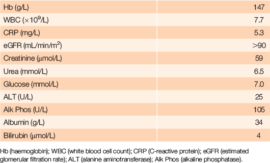
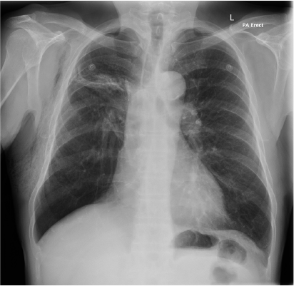
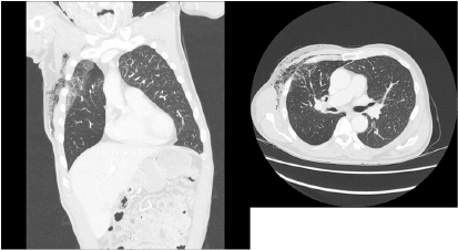
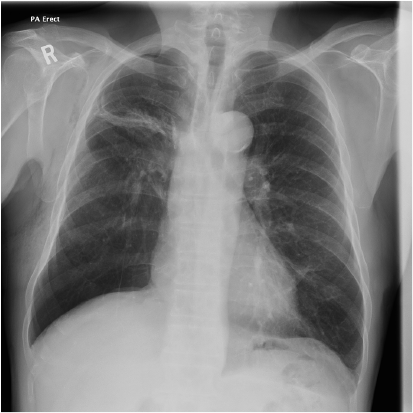
Chest expansion was reduced on the right side and tactile vocal fremitus detected on the right side. Percussion note was hyper-resonant on the right but normal on the left. Auscultation revealed diffuse bilateral rhonchi. Laboratory investigations are shown in Table 1. Chest X-ray demonstrated a small right pneumothorax and right-sided subcutaneous emphysema (Figure 1). A computerised tomography (CT) chest also showed herniation of the right lung through the post-operative defect in the thoracic wall (Figure 2). The patient was initiated on codeine linctus for cough suppression. Over the next 3 days, his symptoms resolved and he remained hemodynamically stable on room air. There was substantial resolution of surgical emphysema (Figure 3). His clinic follow up at 4 weeks showed complete resolution of subcutaneous emphysema.
|
|
|
|
Discussion
Pulmonary herniation is defined as protrusion of lung parenchyma through thoracic wall weakness. Spontaneous pulmonary herniations are rare and more commonly occur in patients with raised intrathoracic pressure such as patients with obesity or COPD.1
Lung herniations can be classified by the anatomic location and the aetiology – congenital or acquired. Approximately 80% of reported cases are acquired and are subcategorized into spontaneous, pathological or traumatic, most being traumatic. They occur more frequently anteriorly in the inferior intercostal spaces because the muscular support is less and the costal cartilage spaces are wider there than in other thoracic wall regions. Patients typically present with chest pain secondary to coughing and a palpable bulge in the chest wall.2
The gold standard management for minimally invasive non-small cell lung cancer is VATS, which is associated with lower post-operative complication rates of pulmonary herniation than traditional thoracotomy.3,4 Fewer than 10 cases of lung herniation have been reported as complications of VATS procedures, with only two known cases reported following VATS lobectomy, both occurring within 1 month of the operation.5,6 Management can be either conservative or surgical. In cases where pain is persistent, operative repair is undertaken to prevent incarceration and strangulation, but no research has investigated long-term treatment outcomes.1 Smoking cessation and weight reduction help decrease the risk of recurrence.2
Our case presented with pulmonary herniation 3 years post VATS procedure. COPD was the added risk factor for this case. We could find no reports of other VATS lobectomy cases where lung herniation presented years after surgery. Moreover, this case is unusual because the patient’s chest pain resolved 1 day after admission allowing his lung herniation to be managed conservatively.
To conclude, lung herniation is a rare presentation with few reported cases in the literature. General practitioners and physicians should keep lung herniation as a differential diagnosis in patients presenting with chest pain secondary to coughing, particularly for people with a history of previous thoracic surgery or trauma.
Competing interests
The authors declare no competing interests.
Funding statement
We did not receive any specific funding for this report.
References
[1] Dahlkemper CL, Greissinger WP. Spontaneous lung herniation after forceful coughing. Am J Emerg Med. 2020; 38 851.e5–6.| Spontaneous lung herniation after forceful coughing.Crossref | GoogleScholarGoogle Scholar |
[2] Scelfo C, Longo C, Aiello M, et al. Pulmonary hernia: Case report and review of the literature. Respirol Case Rep. 2018; 6 e00354
| Pulmonary hernia: Case report and review of the literature.Crossref | GoogleScholarGoogle Scholar | 30302252PubMed |
[3] Jiang Y, Su Z, Liang H, et al. Video-assisted thoracoscopy for lung cancer: who is the future of thoracic surgery? J Thorac Dis. 2020; 12 4427–33.
| Video-assisted thoracoscopy for lung cancer: who is the future of thoracic surgery?Crossref | GoogleScholarGoogle Scholar | 32944356PubMed |
[4] Batıhan G, Yaldız D, Ceylan KC. A rare complication of video-assisted thoracoscopic surgery: lung herniation retrospective case series of three patients and review of the literature. Videosurgery Miniinv. 2020; 15 215–9.
| A rare complication of video-assisted thoracoscopic surgery: lung herniation retrospective case series of three patients and review of the literature.Crossref | GoogleScholarGoogle Scholar |
[5] Ishibashi H, Hirose M, Ohta S. Lung hernia after video-assisted thoracoscopic lobectomy clearly visualized by three-dimensional computed tomography. Eur J Cardiothorac Surg. 2007; 31 938
| Lung hernia after video-assisted thoracoscopic lobectomy clearly visualized by three-dimensional computed tomography.Crossref | GoogleScholarGoogle Scholar | 17337200PubMed |
[6] Johnson C, Weksler B. Lung hernia after video-assisted thoracoscopic lobectomy. Innovations. 2010; 5 300–2.
| Lung hernia after video-assisted thoracoscopic lobectomy.Crossref | GoogleScholarGoogle Scholar | 22437462PubMed |


