Arboviruses in pregnancy: consequences of maternal and fetal infection
William RawlinsonSerology and Virology Division, Department of Microbiology NSW Health Pathology, Prince of Wales Hospital, Sydney, Australia;
School of Women’s and Children’s Health, University of New South Wales; School of Medical Sciences, University of New South Wales;
School of Biotechnology and Biomolecular Sciences, University of New South Wales, Sydney, Australia.
Tel: +61 2 9382 9113, Fax: +61 2 9382 9098, Email: w.rawlinson@unsw.edu.au
Microbiology Australia 39(2) 96-98 https://doi.org/10.1071/MA18028
Published: 6 April 2018
Epidemics and localised outbreaks of infections due to arthropod borne (arbo) viruses, have been described for hundreds of years. Few viruses to date are known to transmit from mother to fetus, causing either teratogenic effects or fetal demise (see recent reviews Charlier et al.1 and Marinho et al.2). Many arboviruses are zoonotic but there appear to be few parallels between the effect of these viruses following human or animal infection during pregnancy. Higher rates of MTCT (mother to child transmission) may be seen (1) where herd immunity is reduced, either because virus is newly introduced into a population (as occurred in Brazil with ZIKV), or where the virus has only recently become endemic (as occurred with West Nile virus (WNV) in the USA in the 1990s), (2) where the arthropod vector is present, (3) where the vector transmits virus efficiently, and (4) in groups of pregnant women exposed, allowing transmission3.
Transmission
There are ~200 million pregnancies annually worldwide, with 90% in regions where arboviruses are endemic: often these are regions with reduced diagnostic capacity4. Arbovirus epidemics have the potential for significant outbreaks of disease in pregnant women and their unborn infants, as evidenced by ZIKV in Brazil where ~17 000 pregnant women had been infected by April 20175. Except for DENV and ZIKV, much is unknown about MTCT of arboviruses, the antenatal and perinatal effects of infection are not well described, and mechanisms of viral teratogenicity are only recognised for some arboviruses1. This means links between adverse pregnancy outcomes and arboviral infections are problematic, particularly where the clinical outcomes are mild, develop over time or are subtle, such as failure to thrive, and mild neurodevelopmental delay.
Effects on the mother
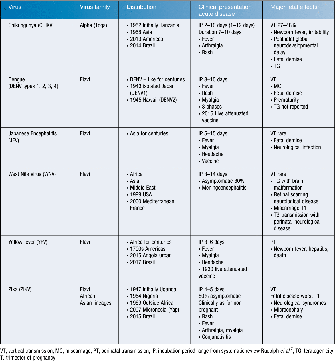
Pregnant women suffer similar symptoms and signs as non-pregnant women, although in general terms, they have a higher risk of more severe effects of viral infection due to the immunosuppression of pregnancy6. The typical syndromes include short incubation time (<1 week), fever, malaise, rash, encephalitis/meningoencephalitis, or haemorrhagic fever (Table 1). Maternal and other infections are asymptomatic in 70–80% of infected individuals, excepting CHIKV (>95% symptomatic often with severe arthritis)8, Yellow fever virus (YFV, ~50% symptomatic)9, and ZIKV Asian lineage (~50% symptomatic with prominent pruritic rash and arthritis)10. Infection in pregnancy with DENV results in increased maternal mortality and severe illness (OR 3.38, 95% CI 2.1–5.42), increased caesarean section and increased postpartum haemorrhages11.

|
Effects on the fetus and newborn
It remains uncertain whether arboviral infection causes fetal damage through direct fetal infection when it occurs, and/or as a result of placental dysfunction causing fetal damage through malnutrition and reduced function, as occurs in other congenital infections12,13. The fetal and neonatal disease caused by arbovirus infection includes fetal demise, premature birth, and neurodevelopmental defects from viral teratogenicity. The latter has been recognised particularly with ZIKV and Venezuelan equine encephalitis (VEE) virus infections, as summarised in Table 1, and in recent reviews1,2. Ross River Virus infection may cause fetal infection, although it is uncertain if fetal disease results14–16.
DENV is the most common arboviral infection globally, and is associated with severe illness in utero, including premature birth (21% versus background rate 11.5%), and miscarriage particularly during the first trimester (T1), and fetal death (13% versus background in utero death rate of 1.8%)11. Maternal symptomatic presentation correlates with rates of fetal demise, with no clear association with serotype. The immune enhancement syndrome seen with second infection from a serotype discordant with a prior DENV serotype infection is also seen in pregnant women, and the fetus17. Other arboviruses appear to less frequently affect the fetus18,19.
The most prominent recent arbovirus-induced fetal malformation has been ZIKV infection, particularly with the Asian lineage in the Americas. This outbreak, initially most severe in Brazil, has been discussed in this journal20, and elsewhere by those at the forefront of epidemic response21–23, and more recently in the United States of America24. It has become clear that regarding ZIKV: (1) infection in pregnant women may result in fetal demise, intrauterine growth restriction, microcephaly, and neurological abnormalities25; (2) disease includes abnormalities of the eye, hearing, craniofacial area, musculoskeletal system; (3) a range of pathological changes occur, with brain abnormalities present in 1–13% of infants of mothers with ZIKV infection; (4) the highest, but not only, risk period is T1, T2; (5) apparently healthy neonates born following maternal ZIKV infection may have longterm neurodevelopmental adverse outcomes; (6) not all outbreaks produce identical phenotypes of fetal and neonatal disease, although infections during pregnancy predominantly result in neurological adverse outcomes of varying severity; (7) screening for ZIKV in pregnant women has been recommended for at-risk populations5; and (8) ZIKV is spread sexually, unusually for an arthropod borne virus, and such spread may result from seminal ZIKV present for up to 120 days in semen, albeit at low titre26.
The diagnosis of arbovirus infections in pregnancy utilises standard molecular methods (predominantly arbovirus specific RT-PCR of blood, cerebrospinal fluid, urine, saliva) and serology (predominantly EIA for detecting virus-specific IgG, IgM), with evidence of seroconversion where specimens are available. There are a range of commercial diagnostic assays available but they vary significantly in their sensitivity and specificity and require care when interpreting results27.
Control of arbovirus infections
Many editorials discuss that recent ZIKV outbreaks highlight the effects of arboviruses on pregnant women, rather than ZIKV being the only cause of virus-induced congenital malformation28. Of the arboviruses known to pose a risk to pregnancy (Table 1), there are vaccines against YFV, JEV and WNV, with vaccines against DENV, CHIKV and ZIKV in various stages of development and testing. Sexual transmission of these viruses (particularly ZIKV) including from asymptomatic hosts, poses a significant challenge. Guidelines are available to inform efforts to minimise this risk. As public health and vaccine efforts are enhanced, it is hoped control of MTCT and neonatal disease from arboviruses will improve.
References
[1] Charlier, C. et al. (2017) Arboviruses and pregnancy: maternal, fetal, and neonatal effects. Lancet Child Adolesc. Health 1, 134–146.| Arboviruses and pregnancy: maternal, fetal, and neonatal effects.Crossref | GoogleScholarGoogle Scholar |
[2] Marinho, P.S. et al. (2017) A review of selected arboviruses during pregnancy. Matern. Health Neonatol. Perinatol. 3, 17.
| A review of selected arboviruses during pregnancy.Crossref | GoogleScholarGoogle Scholar |
[3] Tsai, T.F. (2006) Congenital arboviral infections: something new, something old. Pediatrics 117, 936–939.
| Congenital arboviral infections: something new, something old.Crossref | GoogleScholarGoogle Scholar |
[4] McGready, R. et al. (2010) Arthropod borne disease: the leading cause of fever in pregnancy on the Thai-Burmese border. PLoS Negl. Trop. Dis. 4, e888.
| Arthropod borne disease: the leading cause of fever in pregnancy on the Thai-Burmese border.Crossref | GoogleScholarGoogle Scholar |
[5] Honein, M.A. et al. (2017) Birth defects among fetuses and infants of us women with evidence of possible Zika virus infection during pregnancy. JAMA 317, 59–68.
| Birth defects among fetuses and infants of us women with evidence of possible Zika virus infection during pregnancy.Crossref | GoogleScholarGoogle Scholar |
[6] Scott, G.M. et al. (2012) Cytomegalovirus infection during pregnancy with materno-fetal transmission induces a pro-inflammatory cytokine bias in placenta and amniotic fluid. J. Infect. Dis. 205, 1305–1310.
| Cytomegalovirus infection during pregnancy with materno-fetal transmission induces a pro-inflammatory cytokine bias in placenta and amniotic fluid.Crossref | GoogleScholarGoogle Scholar | 1:CAS:528:DC%2BC38Xksl2rs7o%3D&md5=a74ac71136c83f53f952fdd570ab1a98CAS |
[7] Rudolph, K.E. et al. (2014) Incubation periods of mosquito-borne viral infections: a systematic review Am. J. Trop. Med. Hyg. 90, 882–891.
| Incubation periods of mosquito-borne viral infections: a systematic reviewCrossref | GoogleScholarGoogle Scholar |
[8] Weaver, S.C. and Lecuit, M. (2015) Chikungunya virus and the global spread of a mosquito-borne disease. N. Engl. J. Med. 372, 1231–1239.
| Chikungunya virus and the global spread of a mosquito-borne disease.Crossref | GoogleScholarGoogle Scholar | 1:CAS:528:DC%2BC2MXmtFaiur4%3D&md5=365d76b2e81ae3022d6ac623af396c72CAS |
[9] Cleton, N. et al. (2012) Come fly with me: review of clinically important arboviruses for global travelers. J. Clin. Virol. 55, 191–203.
| Come fly with me: review of clinically important arboviruses for global travelers.Crossref | GoogleScholarGoogle Scholar |
[10] Gallian, P. et al. (2017) Zika virus in asymptomatic blood donors in Martinique. Blood 129, 263–266.
| Zika virus in asymptomatic blood donors in Martinique.Crossref | GoogleScholarGoogle Scholar | 1:CAS:528:DC%2BC2sXhsFSjtLnM&md5=4019342e0d08d9e455831b56f3f071cfCAS |
[11] Machado, C.R. et al. (2013) Is pregnancy associated with severe dengue? A review of data from the Rio de Janeiro surveillance information system. PLoS Negl. Trop. Dis. 7, e2217.
| Is pregnancy associated with severe dengue? A review of data from the Rio de Janeiro surveillance information system.Crossref | GoogleScholarGoogle Scholar | 1:CAS:528:DC%2BC3sXpvVKmsLw%3D&md5=f53491375812f423e429c632397c04fcCAS |
[12] van Zuylen, W.J. et al. (2016) Human cytomegalovirus modulates expression of noncanonical Wnt receptor ROR2 to alter trophoblast migration. J. Virol. 90, 1108–1115.
| Human cytomegalovirus modulates expression of noncanonical Wnt receptor ROR2 to alter trophoblast migration.Crossref | GoogleScholarGoogle Scholar | 1:CAS:528:DC%2BC28Xpt1Omurk%3D&md5=6141ea5b6a844f547d7d60ce99aa9efaCAS |
[13] Maidji, E. et al. (2010) Antibody treatment promotes compensation for human cytomegalovirus-induced pathogenesis and a hypoxia-like condition in placentas with congenital infection. Am. J. Pathol. 177, 1298–1310.
| Antibody treatment promotes compensation for human cytomegalovirus-induced pathogenesis and a hypoxia-like condition in placentas with congenital infection.Crossref | GoogleScholarGoogle Scholar | 1:CAS:528:DC%2BC3cXht1KmsbzK&md5=7effd39db6d5b988ab78ec3f6201cbffCAS |
[14] Aaskov, J.G. et al. (1981)a Evidence for transplacental transmission of Ross River virus in humans. Med. J. Aust. 2, 20–21.
| 1:STN:280:DyaL38%2FhtlWgtA%3D%3D&md5=5c880db5164ba50d834c180b096d7b58CAS |
[15] Aaskov, J.G. et al. (1981)b Effect on mice of infection during pregnancy with three Australian arboviruses. Am. J. Trop. Med. Hyg. 30, 198–203.
| Effect on mice of infection during pregnancy with three Australian arboviruses.Crossref | GoogleScholarGoogle Scholar | 1:STN:280:DyaL3M7lslOmtA%3D%3D&md5=60e3d5eae1049892f3cf835ffe26bd5aCAS |
[16] Aleck, K.A. et al. (1983) Absence of intrauterine infection following Ross River virus infection during pregnancy. Am. J. Trop. Med. Hyg. 32, 618–620.
| Absence of intrauterine infection following Ross River virus infection during pregnancy.Crossref | GoogleScholarGoogle Scholar | 1:STN:280:DyaL3s3ivVOhtA%3D%3D&md5=dae2c6df5667d5465fcf82dfa31e85b9CAS |
[17] Kliks, S.C. et al. (1988) Evidence that maternal dengue antibodies are important in the development of dengue hemorrhagic fever in infants. Am. J. Trop. Med. Hyg. 38, 411–419.
| Evidence that maternal dengue antibodies are important in the development of dengue hemorrhagic fever in infants.Crossref | GoogleScholarGoogle Scholar | 1:STN:280:DyaL1c7otFeksw%3D%3D&md5=6de15587a87405c0a996d5ece68e21fbCAS |
[18] Wenger, F. (1977) Venezuelan equine encephalitis. Teratology 16, 359–362.
| Venezuelan equine encephalitis.Crossref | GoogleScholarGoogle Scholar | 1:STN:280:DyaE1c%2FnsFaltA%3D%3D&md5=803b20b74916550e0a61caf3a485219aCAS |
[19] Torres, J.R. et al. (2016) Congenital and perinatal complications of chikungunya fever: a Latin American experience. Int. J. Infect. Dis. 51, 85–88.
| Congenital and perinatal complications of chikungunya fever: a Latin American experience.Crossref | GoogleScholarGoogle Scholar |
[20] Rawlinson, W. (2016) Pregnancy, the placenta and Zika virus (ZIKV) infection. Microbiol Aust. , 170–174.
[21] Broutet, N. et al. (2016) Zika virus as a cause of neurologic disorders. N. Engl. J. Med. 374, 1506–1509.
| Zika virus as a cause of neurologic disorders.Crossref | GoogleScholarGoogle Scholar | 1:CAS:528:DC%2BC28XhtlOltLjJ&md5=c63e72ac983808423ddd6499e8fee94cCAS |
[22] Calvet, G. et al. (2016) Detection and sequencing of Zika virus from amniotic fluid of fetuses with microcephaly in Brazil: a case study. Lancet Infect. Dis. 16, 653–660.
| Detection and sequencing of Zika virus from amniotic fluid of fetuses with microcephaly in Brazil: a case study.Crossref | GoogleScholarGoogle Scholar |
[23] Brasil, P. et al. (2016) Zika virus infection in pregnant women in Rio de Janeiro—preliminary report. N. Engl. J. Med. 375, 2321–2334.
| Zika virus infection in pregnant women in Rio de Janeiro—preliminary report.Crossref | GoogleScholarGoogle Scholar |
[24] Fitzgerald, B. et al. (2018) Birth defects potentially related to Zika virus infection during pregnancy in the United States. JAMA 25 January.
[25] Moura da Silva, A.A. et al. (2016) Early growth and neurologic outcomes of infants with probable congenital Zika virus syndrome. Emerg. Infect. Dis. 22, 1953–1956.
| Early growth and neurologic outcomes of infants with probable congenital Zika virus syndrome.Crossref | GoogleScholarGoogle Scholar |
[26] de Laval, F.D. et al. (2017) Kinetics of Zika viral load in semen. N. Engl. J. Med. 377, 697–699.
| Kinetics of Zika viral load in semen.Crossref | GoogleScholarGoogle Scholar |
[27] Bingham, A.M. et al. (2016) Comparison of test results for Zika virus RNA in urine, serum, and saliva specimens from persons with travel-associated Zika virus disease—Florida. MMWR Morb. Mortal. Wkly. Rep. 65, 475–478.
| Comparison of test results for Zika virus RNA in urine, serum, and saliva specimens from persons with travel-associated Zika virus disease—Florida.Crossref | GoogleScholarGoogle Scholar |
[28] Kourtis, A.P. et al. (2014) Pregnancy and infection. N. Engl. J. Med. 371, 1077.
Biography
Professor William Rawlinson AM FAHMS is a Medical Virologist and is Director of the Division of Serology and Virology (SAViD) and a NSW State Reference Laboratory in HIV, in the Department of Microbiology SEALS. He is a consultant position to the Department of Infectious Diseases, Prince of Wales and Sydney Children’s Hospital. He holds a conjoint academic position as Professor in the School of Medical Science and the School of Biotechnology and Biomolecular Sciences at The University of New South Wales. The research group that he heads studies congenital infections, enteroviruses, hepatitis viruses, respiratory viruses, novel antivirals and antiviral resistance.


