

Use of a smartphone-based, non-mydriatic fundus camera for patients with red flag ophthalmic presentations in a rural general practice
Scott Davidson 1 * , Waldir Rodrigues de Souza Jr 2 3 , Kyle Eggleton 4
1
2
3
4
Abstract
Fundus examination by direct ophthalmoscopy is widely used in general practice; however, it offers limited field of view, requires close approximation to the patient, has a steep learning curve and is a difficult skill to master and maintain. Non-mydriatic fundus photography (NMFP) offers an alternative with a wider field of view, ability for image analysis and transmission, and is able to be conducted by allied healthcare staff.
This study aimed to compare the use of direct ophthalmoscopy with smart-phone NMFP in a large rural general practice. It also aimed to analyse the number of adequate views and positive findings achieved with each instrument and the impact of NMFP on ophthalmology referral decisions.
Patients aged ≥16 years presenting to Dargaville Medical Centre (Dargaville, New Zealand) with visual disturbance, headache, hypertensive urgency (systolic blood pressure (BP) >200 mmHg or diastolic BP >120 mmHg), transient ischemic attack (TIA) or stroke were enrolled prospectively into an observational study of visualisation, diagnosis and management impact for a 1-year period (n = 152, 304 eyes). Direct ophthalmoscopy findings and management plans were documented by the attending general practitioner (GP), and then again following assessment of the NMFP.
NMFP significantly improved visualisation of the fundal structures with an increase in adequate views achieved of both the optic disc and the retina. Inter-rater agreement between the referring GP and ophthalmologist was good.
The use of NMFP in general practice might result in greater accuracy in diagnosing retina and optic disc disease. Routine transmission of NMFP images to specialist eye clinics as part of the referral might improve management and result in health system efficiencies.
Keywords: direct ophthalmoscope, fundoscopy, fundus photo, ophthalmology, optic nerve, retina, rural, general practice.
| WHAT GAP THIS FILLS |
| What is already known: General practitioners find examining the retina with direct ophthalmoscopy challenging and have low confidence in interpreting findings. |
| What this study adds: General practitioners using smart-phone non-mydriatic fundus photography achieved more adequate views and positive findings of the optic nerve and fundus compared to direct ophthalmoscopy and had close agreement for management referral with the reviewing ophthalmologist. |
Introduction
Eye conditions presenting to general practitioners (GP) are common, accounting for 2.2 consultations per 100.1 However, GPs often find ophthalmology challenging, with 27–31% of presentations being referred.1,2 Of those patients acutely referred to specialist eye clinics, only 36–57% have a correct diagnosis made by a GP.3,4 One technological solution to improving ophthalmologic diagnosis is non-mydriatic fundus photography (NMFP). These images can be taken by allied healthcare staff, offer a wide field of view and can be electronically transmitted for review. In this study, the use of NMFP in a large rural general practice is evaluated to examine the impact on management of patients presenting with ophthalmologic red flag symptoms.
Diagnosis and management of patients presenting to general practice with red flag symptoms, such as blurred vision or headache generally require good ophthalmic skills, including fundoscopy. GPs, however, receive limited ophthalmology training during medical school. Generally, 1–2 weeks of training is reported by medical students and junior medical officers in North America, Australasia and the UK.5–9 A recent New Zealand (NZ) medical student survey demonstrated low confidence in ophthalmic skills and knowledge.5 GPs report that direct ophthalmoscopy is an important or essential skill; however, it is one of the more difficult to master,6,7,10,11 resulting in its underuse in practice.12–14
A survey of UK GPs revealed that 43% lacked confidence in using a direct ophthalmoscope.15 Another study showed that only a low number of GP referrals (4%) to specialist eye clinics, reported fundoscopy findings.16 In GP referrals relating to cataracts, up to 15% had undiagnosed retinal problems.17 Concordance with ophthalmologist diagnosis, in this particular study, was only 68% of referrals with a retinal diagnosis. An inability to view the fundus by GPs has led to misdiagnosis of optic neuropathy, papilloedema, hypertensive retinopathy, retinal haemorrhage and detachment, with adverse outcomes reported for patients.4,18
One method of improving diagnosis is the use of NMFP. NMFP uses indirect ophthalmoscopy to view an inverted real image of the fundus created by a condensing lens and coaxial light source. A dark room is used, and the camera uses an infrared light source and fixation spot to focus the image of the retina before a white flash is activated to obtain a colour photograph of the retina. The camera may be handheld or mounted with the patients head in a head rest similar to a slit lamp microscope.19
Use of NMFP has been shown to offer higher detection of fundus pathology in emergency department and inpatient settings.20–22 There has been investigation of its use in general practice for diabetes retinal screening; however, to our knowledge, it has not been studied in the diagnosis of patients with red flag ophthalmic symptoms in general practice and may offer advantages over direct ophthalmoscopy.
Methods
The setting of the study was the Dargaville Medical Centre (DMC), Dargaville, New Zealand, a single large rural group general practice in Northland’s western Kaipara area, with approximately 12,159 enrolled patients (European 62.2%, Māori 31.4%, Pacific peoples 2.6%). The nearest ophthalmology and optometric services are 60 km away in Whangarei. The lead author is a GP at DMC. The study enrolled patients from 15 November 2021 to 23 November 2022 and was a prospective, observational study of standard care with direct ophthalmoscopy compared to standard care plus NMPF for diagnosis, referral, management impact and GP inter-rater agreement with specialist care.
Eligible participants in the study were ambulatory patients aged ≥16 years presenting to DMC with visual disturbance, headache, hypertensive urgency (systolic blood pressure (BP) >200 mmHg or diastolic BP >120 mmHg), TIA or stroke. Exclusion criteria were non-ambulatory patients, patients in need of resuscitation or patients deemed too unwell by the treating team.
Following standard care, eligible patients were provided with an information sheet and written consent form. The study involved undertaking direct ophthalmoscopy followed by fundal photographs of one or both of the patient’s eyes. The study protocol is outlined in more detail in the Supplementary material. The consulting GP filled in the study data sheet recording clinical details including adequate view assessment, direct ophthalmoscopy findings, diagnosis and the patient management plan before viewing the fundal photographs and then revising the findings, diagnosis and management. Urgent acute presentations were referred by phone to Whangarei Hospital Eye Clinic, a secondary care service, or to the Greenlane Eye Centre Auckland, a tertiary care service.
The anonymised retinal photographs and clinical information were recorded on a secure online platform and reviewed by an ophthalmologist. Suggested changes to the management plan were sent to DMC.
No formal training was given in fundoscopy to the GPs in the study; however, the lead author has a special interest in ophthalmology and works as a GP with special interest (GPSI) in the Whangarei Hospital Eye Clinic. Recruitment and study protocol was discussed at a 15-min weekly practice team meeting attended by all practice staff.
A two-sample comparison of proportions power calculation was carried out using R-Studio (Posit, Boston, USA). An assumed positive abnormal fundus photograph rate of 0.06 (a conservative estimate compared to other emergency department (ED) studies),21,22 and a significance level of 0.05 with 126 patients was required to achieve a power >80%.
Data were analysed by descriptive statistics, and adequate views and positive finding detection rates (PFDR) were calculated for GPs using direct ophthalmoscopy and NMFP and compared using rate ratios. Inter-rater kappa coefficients were calculated to assess agreeability between GP and ophthalmologist assessment of photos.
Ethics
Consultation with local the Māori Health provider, Te Ha Oranga, and local kaumātua was undertaken to ensure engagement of Māori patients, with suggestions incorporated into the study design. Participants were offered karakia provided by kaumātua. Consultations were free for participants. Ethics approval was obtained from the New Zealand Health and Disability Ethics Committee (ethics number – HDEC 21/NTB/233).
Results
In total, 152 participants (304 eyes) were recruited into the study, which resulted in a power of 86.94% to detect an abnormal fundus.
Characteristics of participants are presented in Table 1.
| Variable | n (%)A | |
|---|---|---|
| Age (mean) | 64 years | |
| Sex | ||
| Female | 75 (49.3) | |
| Male | 77 (50.7) | |
| Ethnicity | ||
| European | 108 (71) | |
| Māori | 38 (25) | |
| Pacific peoples | 3 (2) | |
| Asian | 1 (0.7) | |
| Other | 2 (1.3) | |
| Average visual acuity | 6/13.6 (mean logMAR 0.3235 + −0.4182) | |
| Presenting complaints | ||
| Visual disturbance | 127 (84) | |
| Headache | 35 (22) | |
| Hypertensive urgency | 5 (3) | |
| Neurological deficit (transient ischemic attack / cerebrovascular accident) symptoms | 3 (2) | |
GPs using NMFP had a higher number of adequate views, positive and normal findings for the optic disc and the retina.
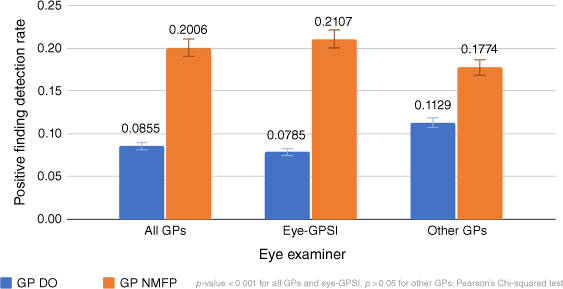
The positive finding detection rate (PFDR) of acute and chronic abnormalities was the number of positive findings per number of eyes examined (combining views of disc and retina), as detailed in Tables 2 and 3. The use of NMFP, when compared with direct ophthalmoscopy, significantly increased the PFDR, from 0.0855 to 0.2039 (Fig. 1); however, it only marginally increased the number of referrals (114 compared to 113).
| Optic disc | Retina | ||||||
|---|---|---|---|---|---|---|---|
| Direct ophthalmoscopy | NMFP | % change | Direct ophthalmoscopy | NMFP | % change | ||
| Adequate views per eye examined | 236 | 273 | 15.6 | 151 | 270 | 78.8 | |
| Inadequate views per eye examined | 68 | 25 | −63.2 | 153 | 24 | −84.32 | |
| Positive findings per eye examined | 12 | 17 | 41.6 | 14 | 48 | 242.8 | |
| Normal findings per eye examined | 224 | 256 | 14.2 | 137 | 222 | 62 | |
| Non-mydriatic fundus photography – Ophthalmologist | |||||
|---|---|---|---|---|---|
| Non-mydriatic fundus photography – General Practitioner | No referral | Referral | Total | ||
| No referral | 52 | 8 | 60 | ||
| Referral | 3 | 89 | 92 | ||
| Total | 55 | 97 | 152 | ||
Pearson’s Chi-squared test, P-value < 0.001.
The inter-rater agreement (kappa coefficient) between GP and ophthalmologist assessment for NMFP was 0.84632, which is considered a very good agreement.23 The inter-rater agreement between GP and ophthalmologist, when GPSI results were excluded was 0.73617, which is also considered a good agreement (Table 3).23
Following ophthalmologist review, with mydriatic and NMFPs, 20 patient referral decisions were recommended for alternative management. Ten patients that had been referred to ophthalmology were thought to be able to be managed in general practice. Seven patients remaining under GP care were recommended GPSI or ophthalmology referral, and two optometry referrals and one patient referred to ophthalmology were redirected to a vitreoretinal service.
Following review with mydriatic and NMFP by the attending GP, nine referral decisions were changed. Two were re-directed from secondary care ophthalmology to a tertiary vitreoretinal service for retinal detachments, two back to GP care from ophthalmology or optometry referral, three from GP to ophthalmology care, one from ophthalmology to neurology and one from GP to general medicine.
GPs reported NMFP as helpful in 134 of 152 patient cases. Abnormal fundus findings included: retinal detachment, branch and central retinal vein occlusion (see Supplementary Fig. S1), branch retinal artery occlusion, age-related macular degeneration haemorrhage and dry, bilateral disc pallor, central serous retinopathy, toxoplasmosis vitritis/retinitis (see Supplementary Fig. S2), macular scar/dislocated intraocular lens, glaucomatous disc cupping, diabetic retinopathy, hydroxychloroquine maculopathy and retinal haemorrhage.
Discussion
The findings from this study show that GP NMFP can decrease the uncertainty of fundal and disc examination over direct ophthalmoscopy. In addition, GP NMFP has high agreement with ophthalmologists examining fundal photographs for detecting abnormal retinal findings and referral decisions and is able to be performed by non-physician members of the primary care team.
Numerous barriers to the use of direct ophthalmoscopy have led to an interest in fundus photography for non-ophthalmologists in evaluating the retina.14,24,25 Medical students have reported a preference for retinal photography over direct ophthalmoscopy and had more accuracy in identifying fundus pathology.26 NMFP has been shown to have higher sensitivity at detecting diabetic retinopathy compared to direct ophthalmoscopy.27 In addition, NMFP has been demonstrated to be superior to direct ophthalmoscopy in diagnostic accuracy and impact on management decisions in patients presenting with visual disturbance, headache, hypertensive urgency or neurological symptoms compared to direct ophthalmoscopy.21,22
Studies, however, show variable ability of GPs in reporting and grading fundus photographs.28,29 In one study, GPs had only fair agreement compared to trained non-physician graders and ophthalmologists for the grading of NMFPs for diabetic retinopathy.29 In another study, comparing diagnostic accuracy using stereoscopic fundal photographs in recognising papilloedema, GPs had a sensitivity of 84.5% and specificity of 59.3% compared to neuro-ophthalmologists.30 In contrast, this study shows very good inter-rater agreement between GPs and ophthalmologists; however, 80% of participants were seen by the GPSI, which would be expected in terms of an increased agreement with ophthalmologists compared to the remaining GPs with less training and experience.
In rural areas where often no optometric or ophthalmology service is locally available, ability to use NMFP for tele-ophthalmology assessment, triage and referral offers potential improvement in GP diagnostic ability, health system efficiency, patient care and potential travel savings. Although in our study there was little difference in the number of patients referred to ophthalmology, for a low number, referral was able to be made to a tertiary service for retinal detachment repair and retrieval of dislocated intra-ocular lens. Also in our study following NMFP review by an ophthalmologist, a number of patients were returned to GP care with advice. This demonstrates a potential to decrease the demand on ophthalmology services. In an Australian study of a tele-ophthalmology service, patients were less likely to require follow-up by an eye care provider if images were provided by the primary care provider.31
Use of smartphone-based fundoscopy has enabled the development of lower cost fundus cameras. Many, however, have required mydriasis to achieve satisfactory results.32 One NZ general practice-based study, which used mydriasis and smartphone fundoscopy in patients presenting with visual complaints, demonstrated 94.5% of photos being of acceptable, good or very good quality for detecting optic disc abnormalities, but only 50% for detecting macular abnormalities.24 Our study that used a non-mydriatic smartphone-based camera had similar success at imaging the optic nerve, but was significantly better at imaging the macula.
Our study is limited by being a single-centre study. A further multicentred study with a wider range of GPs with different ophthalmology experience would be beneficial. A strength of the study was the duration of the study over a year, which avoided seasonal bias in presentation and a comparable number of enrolled patients to other studies, therefore enabling statically significant results. A weakness that may have impacted enrolled numbers was its implementation during the COVID-19 pandemic level two measures, with limited numbers of patients attending face-to-face consultations.
Conclusion
In our study, GPs examining the eye using NMFP achieved a higher PFDR compared to when direct ophthalmoscopy is used, and had overall good inter-rater agreement with the reviewing ophthalmologist. The development of a smartphone-based, non-mydriatic fundus camera offers a lower cost alternative to acquire wide angle images of the retina that can be easily and securely transmitted. It provides the examining doctor with a good view of the retina and the ability to transmit the image for referral, triage or acute advice. Implementing teleophthalmology care into rural and remote areas may improve health system efficiency.
Data availability
The data that support this study will be shared upon reasonable request to the corresponding author.
Conflicts of interest
ODOCS Eye Care, Dunedin, Otago provided non-financial support and the nun IR smartphone fundus camera for this study.
Declaration of funding
The New Zealand Rural GP Network; New Zealand Institute of Rural Health and the Charitable Education and Research Trust, Auckland and Northland Faculties, Royal New Zealand College of General Practitioners provided funding for this study.
References
1 Morgan S, Tapley A, Henderson KM, et al. Australian general practice trainees’ exposure to ophthalmic problems and implications for training: a cross-sectional analysis. J Prim Health Care 2016; 8(4): 295-302.
| Crossref | Google Scholar | PubMed |
3 Perumal D, Niederer R, Raynel S, et al. Patterns of ophthalmic referral and emergency presentations to an acute tertiary eye service in New Zealand. N Z Med J 2011; 124(1340): 35-47.
| Google Scholar |
4 Statham MO, Sharma A, Pane AR. Misdiagnosis of acute eye diseases by primary health care providers: incidence and implications. Med J Aust 2008; 189(7): 402-4.
| Crossref | Google Scholar | PubMed |
5 Han L, Ogbuehi KC. Focus on undergraduate ophthalmology teaching, survey of final year medical students in a New Zealand medical school. Clin Exp Ophthalmol 2020; 48(7): 1001-2.
| Crossref | Google Scholar | PubMed |
6 Succar T, McCluskey P, Grigg J. Enhancing medical student education by implementing a competency-based ophthalmology curriculum. Asia Pac J Ophthalmol 2017; 6(1): 59-63.
| Crossref | Google Scholar | PubMed |
7 Gelston CD, Patnaik JL. Ophthalmology training and competency levels in caring for patients with ophthalmic complaints among United States internal medicine, emergency medicine, and family medicine residents. J Educ Eval Health Prof 2019; 16: 25.
| Crossref | Google Scholar | PubMed |
8 Noble J, Somal K, Gill HS, et al. An analysis of undergraduate ophthalmology training in Canada. Can J Ophthalmol 2009; 44(5): 513-8.
| Crossref | Google Scholar | PubMed |
9 Baylis O, Murray PI, Dayan M. Undergraduate ophthalmology education – A survey of UK medical schools. Med Teach 2011; 33(6): 468-71.
| Crossref | Google Scholar | PubMed |
10 Zhang HH, Hepschke JL, Shulruf B, et al. Sharpening the focus on ophthalmology teaching: perceptions of medical students and junior medical officers. Clin Exp Ophthalmol 2018; 46(9): 984-93.
| Crossref | Google Scholar | PubMed |
11 Ah-Chan JJ, Sanderson G, Vote BJ, et al. Undergraduate ophthalmology education survey of New Zealand ophthalmologists, general practitioners and optometrists. Clin Exp Ophthalmol 2001; 29(6): 416-25.
| Crossref | Google Scholar | PubMed |
12 Jackson C, De Jong I, Glasson W. Royal Australian College of Ophthalmologists and Royal Australian College of General Practitioners National GP eye skills workshops: Colleges and divisions reskilling general practice. Clin Exp Ophthalmol 2000; 28(5): 347-9.
| Crossref | Google Scholar | PubMed |
13 Lamirel C, Bruce BB, Wright DW, et al. Quality of nonmydriatic digital fundus photography obtained by nurse practitioners in the emergency department: the FOTO-ED study. Ophthalmology 2012; 119(3): 617-24.
| Crossref | Google Scholar | PubMed |
14 Mackay DD, Garza PS, Bruce BB, et al. The demise of direct ophthalmoscopy: a modern clinical challenge. Neurol Clin Pract 2015; 5(2): 150-7.
| Crossref | Google Scholar | PubMed |
15 Shuttleworth GN, Marsh GW. How effective is undergraduate and postgraduate teaching in ophthalmology? Eye 1997; 11(5): 744-50.
| Crossref | Google Scholar | PubMed |
16 Pierscionek TJ, Moore JE, Pierscionek BK. Referrals to ophthalmology: optometric and general practice comparison. Ophthalmic Physiol Opt 2009; 29(1): 32-40.
| Crossref | Google Scholar | PubMed |
17 Davey CJ, Green C, Elliott DB. Assessment of referrals to the hospital eye service by optometrists and GPs in Bradford and Airedale. Ophthalmic Physiol Opt 2011; 31(1): 23-8.
| Crossref | Google Scholar | PubMed |
18 Stunkel L, Sharma RA, Mackay DD, et al. Patient harm due to diagnostic error of neuro-ophthalmologic conditions. Ophthalmology 2021; 128(9): 1356-62.
| Crossref | Google Scholar | PubMed |
19 Mackay DD, Bruce BB. Non-mydriatic fundus photography: a practical review for the neurologist. Pract Neurol 2016; 16: 343-51.
| Crossref | Google Scholar | PubMed |
20 He G, Dunn HP, Ahmad KE, et al. Fundoscopy use in neurology departments and the utility of smartphone photography: a prospective prevalence and crossover diagnostic accuracy study amongst neurology inpatients. Eur J Neurol 2022; 29(8): 2463-2472.
| Crossref | Google Scholar | PubMed |
21 Bruce BB, Thulasi P, Fraser CL, et al. Diagnostic accuracy and use of nonmydriatic ocular fundus photography by emergency physicians: phase II of the FOTO-ED study. Ann Emerg Med 2013; 62(1): 28-33.
| Crossref | Google Scholar | PubMed |
22 Dunn HP, Browning SD, Thomson D, et al. Impact on patient management of non‐mydriatic fundus photography compared to direct ophthalmoscopy in a regional Australian emergency department. Emerg Med Australas 2022; 34(2): 186-93.
| Crossref | Google Scholar | PubMed |
24 Singh A, Cheyne K, Wilson G, et al. On the use of a new monocular-indirect ophthalmoscope for retinal photography in a primary care setting. N Z Med J 2020; 133(1512): 31-8.
| Google Scholar | PubMed |
25 Dunn HP, Kang CJ, Marks S, et al. Optimising fundoscopy practices across the medical spectrum: a focus group study. PLoS One 2023; 18(1): e0280937.
| Crossref | Google Scholar | PubMed |
26 Kelly LP, Garza PS, Bruce BB, et al. Teaching ophthalmoscopy to medical students (the TOTeMS study). Am J Ophthalmol 2013; 156(5): 1056-61.
| Crossref | Google Scholar |
27 Siu SC, Ko TC, Wong KW, et al. Effectiveness of non-mydriatic retinal photography and direct ophthalmoscopy in detecting diabetic retinopathy. Hong Kong Med J 1998; 4: 367-70.
| Google Scholar | PubMed |
28 Castro AF, Silva-Turnes JC, Gonzalez F. Evaluation of retinal digital images by a general practitioner. Telemed J E Health 2007; 13(3): 287-92.
| Crossref | Google Scholar | PubMed |
29 Bhargava M, Cheung CY, Sabanayagam C, et al. Accuracy of diabetic retinopathy screening by trained non-physician graders using non-mydriatic fundus camera. Singapore Med J 2012; 53(11): 715-9.
| Google Scholar | PubMed |
30 Johnson LN, Hepler RS, Bartholomew MJ. Accuracy of papilledema and pseudopapilledema detection: a multispecialty study. J Fam Nurs 1991; 33(4): 381-6.
| Google Scholar | PubMed |
31 Johnson KA, Meyer J, Yazar S, et al. Real‐time teleophthalmology in rural Western Australia. Aust J Rural Health 2015; 23(3): 142-9.
| Crossref | Google Scholar | PubMed |
32 Wintergerst MWM, Jansen LG, Holz FG, et al. Smartphone-based fundus imaging–Where are we now? Asia Pac J Ophthalmol 2020; 9(4): 308-14.
| Crossref | Google Scholar | PubMed |