

Complete sigmoid colon erosion by an intrauterine contraception device in an ectopic pregnancy: a case report
Chuah Jun Sen 1 2 3 , Nur Hayati Abd Samad 2 , Tan Jih Huei 2 , Lee Ee Peng 21 Department of General Surgery, Pusat Perubatan Universiti Kebangsaan Malaysia, Cheras, Malaysia.
2 Department of General Surgery, Hospital Sultanah Aminah, Ministry of Health Malaysia, Johor Bahru, Malaysia.
3 Corresponding author. Email: Vincentchuahjs@gmail.com
Journal of Primary Health Care 13(3) 283-286 https://doi.org/10.1071/HC21084
Published: 23 September 2021
Journal Compilation © Royal New Zealand College of General Practitioners 2021 This is an open access article licensed under a Creative Commons Attribution-NonCommercial-NoDerivatives 4.0 International License
Abstract
INTRODUCTION: An intrauterine contraceptive device (IUCD) is a common contraception method used for family planning. IUCD erosion into adjacent organs is a rare but serious complication of IUCD use.
CASE PRESENTATION: A 41-year-old female presented to us with a leaking left ectopic pregnancy. Emergency laparotomy and left salpingectomy were performed. A copper ICUD was found intraperitoneally and part of it had completely eroded into the sigmoid colon. Sigmoid colotomy was performed and the IUCD was removed successfully. Further history revealed that the patient had her IUCD inserted 12 years previously but was forgotten. The patient was discharged well after 4 days of admission.
DISCUSSION: Erosion of an IUCD into the colon is uncommon and may be asymptomatic or present with bowel perforation and obstruction. There should be a high index of suspicion for pregnancy occurring among women post-IUCD insertion. A misplaced IUCD can cause chronic inflammation of the fallopian tube, which may alter tubal functionality and increase the risk of ectopic pregnancy. Family planning is commonly done in primary health care. Primary care education and counselling are essential to improve awareness of fertile women to prevent similar complications. Periodic examination of IUCD string either by users or primary health-care practitioners is crucial. Ultrasound can be advocated if there are difficulties with the insertion. An abdominal radiograph is useful and should be performed in the case of missing IUCDs.
KEYwords: Intrauterine device; intestinal perforation; ectopic pregnancy; primary health care.
| WHAT GAP THIS FILLS |
| What is already known: IUCDs are a common contraception method used for family planning. IUCD erosion to other organs is uncommon, but is a potentially serious complication. |
| What this case report adds: There should be a high index of suspicion of IUCD misplacement in pregnancy, which occurs among females with post-IUCD insertion. A forgotten intraperitoneal IUCD may potentially increase risk of ectopic pregnancy due to chronic inflammation of the fallopian tube. Primary health-care education, counselling and periodic examination of IUCD string are essential to improve the awareness of fertile patients to prevent similar complications. An abdominal radiograph is valuable and should be performed in the case of a missing IUCD. |
Introduction
Intrauterine contraceptive devices (IUCDs) are commonly used as an effective long-term reversible contraception method. These devices can easily be inserted, but they are not without potential complications. They can migrate to other abdominal organs causing disastrous complications.1 Eroding into the bowel may result in bowel perforation, intraperitoneal abscess formation or active bleeding. We report an unusual case of complete sigmoid colon erosion by an IUCD in a patient concurrently having a leaking ectopic pregnancy. The challenges in diagnosis and multidisciplinary surgical management are discussed.
Case presentation
A 41-year-old female with a 4-week period of amenorrhoea presented with lower abdominal pain for 2 days. She was gravida 9 and para 4 with a history of four miscarriages. Physical examination revealed a tender left lower abdomen with voluntary guarding. A urine pregnancy test was positive for pregnancy and blood investigation showed leucocytosis with a white cell count of 22 × 11.0 × 109/L. Abdominal and transvaginal ultrasound scans revealed a left adnexal mass with presence of minimal free fluid. A diagnosis of leaking left ectopic pregnancy was suspected. An emergency laparotomy was performed.
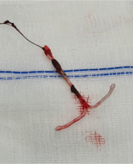
Intraoperatively, there was a left tubal pregnancy measuring ~4 × 3 cm. A copper IUCD was found intraperitoneally and part of its horizontal arm had eroded into the mesenteric border of the sigmoid colon (Fig. 1). There was a dense adhesion around the copper IUCD, sigmoid colon, posterior surface of the uterus and left fallopian tube. After careful adhesiolysis, left salpingectomy was performed. The copper IUCD had completely eroded into the mucosal layer of the sigmoid colon. Fortunately, there was no faecal contamination. Sigmoid colotomy was performed, and the copper IUCD was removed as a whole (Fig. 2). Subsequently, the colotomy site was closed primarily using polyglyconate 2/0. Right salpingectomy was conducted as requested for sterilization.

|
|
Postoperatively, the patient recalled that she had a copper IUCD inserted 12 years previously, after her second pregnancy. Within the following 5 years, she had two subsequent vaginal deliveries. She thought the copper IUCD was expelled spontaneously but did not confirm this with a medical practitioner. The IUCD was left forgotten. She later complained of chronic intermittent abdominal pain and discomfort for years. She was discharged well after 4 days of admission and was asymptomatic at her third month follow up.
Discussion
A misplaced IUCD is most commonly found in the omentum (26.7%). Involvement of the colon is uncommon.2 It may be asymptomatic or present with bowel perforation and obstruction.3 In the current case, the IUCD was only detected intraoperatively. From this case, we learned that it might be worthwhile to screen women with missing IUCDs with an abdominal radiograph, despite the performance of a gynaecology ultrasonography assessment. An abdominal radiograph is reported as the first imaging modality to locate an IUCD and it has high specificity and sensitivity for this purpose.4
The absolute risk of ectopic pregnancy is lower for women with an IUCD, but not for a misplaced device.5 An IUCD in the peritoneal cavity acted as a foreign body, causing local inflammation to the surrounding tissue, including the fallopian tube. Chronic inflammation of the fallopian tube may alter tubal functionality and cause pelvic adhesive disease,6 which increases the risk of ectopic pregnancy. This patient presented with ectopic pregnancy, but we did not expect to find a forgotten IUCD, which is possibly the culprit for the tubal dysfunction and resultant ectopic pregnancy.
As increasing numbers of misplaced IUCDs have been reported and 2,3 primary health-care practitioners and gynaecologists should have heightened awareness of this complication. The benefits of IUCD are unquestionable despite the uncommon risk of misplacement or uterine perforation. A possible reason for IUCD misplacement is thought due to uterine perforation. It is believed that uterine perforation occurs mainly during its insertion,2 but it may also occur spontaneously through the gradual erosion of the uterine wall as a chronic inflammatory reaction.7 A correct IUCD insertion technique by an experienced or well-trained health-care practitioner is crucial in preventing complications from arising. Ultrasound can be advocated to assure proper IUCD insertion and allow early identification of uterine perforation.1 Patients need adequate and reinforced, counselling and education regarding maintenance and complications of their IUCD. Education helps to reduce the chance of IUCDs being left forgotten. Periodic examination of the IUCD by users or health-care practitioners is crucial to ensure the IUCD remains in situ. It can be conducted either by checking for the string in the cervical opening or using ultrasound.
Although an IUCD embedded in the colon wall can be removed endoscopically,8 we removed the IUCD with a colotomy, as this is technically more straightforward when the abdomen is already accessed in response to a leaking ectopic pregnancy. If an IUCD is found incidentally, endoscopic retrieval may be attempted, but this may pose a risk of colon perforation, as part of the IUCD may be deeply embedded.2,3,7
Conclusion
There is an increasing number of reports related to IUCD misplacement or forgotten IUCDs. Primary health-care education and counselling are essential to improve the awareness of fertile patients to prevent similar complications. Provision of periodic examination of the IUCD string by users, primary health-care practitioners or gynaecologists is encouraged. Ultrasound is advocated if there are any difficulties with the insertion. An abdominal radiograph is valuable and should be performed in the case of missing IUCD.
Consent
Written informed consent was obtained from the patient for publication of this case report and accompanying images.
Competing interests
The authors have no competing interests to declare.
Declaration of funding
This paper did not receive any specific funding.
Data availability statement
Data are made available in a public repository and can be accessed via a DOI.
Author contributions
Conceptualization: CJS. Data curation: CJS, Nurhayati, TJH. Formal analysis: CJS, Nurhayati, TJH. Methodology: CJS, Nurhayati, TJH, LEP. Project administration: CJS, TJH, LEP. Visualisation: CJS, LEP. Writing original draft: CJS. Writing review and editing: CJS, Nurhayati, TJH, LEP.
Acknowledgements
This report did not require approval from an institutional ethical review board. All authors agreed with the content of the final manuscript. The authors conformed to the provisions of the Declaration of Helsinki in 1995 (as revised in Brazil in 2013).
References
[1] Tan JH, Lip H, Ong W, Omar S. Intrauterine contraceptive device embedded in bladder wall with calculus formation removed successfully with open surgery. Malays Fam Physician. 2019; 14 29–31.| 31827733PubMed |
[2] Gill RS, Mok D, Hudson M, et al. Laparoscopic removal of an Intra-Abdominal Intrauterine Device: case and systematic review. Contraception. 2012; 85 15–8.
| Laparoscopic removal of an Intra-Abdominal Intrauterine Device: case and systematic review.Crossref | GoogleScholarGoogle Scholar | 22067801PubMed |
[3] Johri V, Vyas KC. Misplaced intrauterine contraceptive devices: common errors, uncommon complications. J Clin Diagn Res. 2013; 7 905–7.
| Misplaced intrauterine contraceptive devices: common errors, uncommon complications.Crossref | GoogleScholarGoogle Scholar | 23814739PubMed |
[4] Goswami D, Ravi AK, Sharma A. Missing IUCD strings: role of imaging in locating the misplaced device. J Clin Diagn Res. 2017; 11 QJ01–02.
| Missing IUCD strings: role of imaging in locating the misplaced device.Crossref | GoogleScholarGoogle Scholar | 28571217PubMed |
[5] Furlong LA. Ectopic pregnancy risk when contraception fails. A review. J Reprod Med. 2002; 47 881–5.
| 12497674PubMed |
[6] Adoni A, Ben Chetrit A. The management of intrauterine devices following uterine perforation. Contraception. 1991; 43 77–81.
| The management of intrauterine devices following uterine perforation.Crossref | GoogleScholarGoogle Scholar | 1825971PubMed |
[7] Turner A. Intrauterine device migration. Emerg Med. 2016; 48 417–20.
| Intrauterine device migration.Crossref | GoogleScholarGoogle Scholar |
[8] Chandrasekar TS, Gokul BJ, Yogesh KR, et al. A new endoscopic method of retrieval of a migrated and transmurally embedded intrauterine contraceptive device in the rectum. Indian J Gastroenterol. 2016; 35 489–91.
| A new endoscopic method of retrieval of a migrated and transmurally embedded intrauterine contraceptive device in the rectum.Crossref | GoogleScholarGoogle Scholar | 27796939PubMed |