Macroscopic and dermoscopic evaluation used to differentiate subungual haemorrhage from melanocytic lesions
Mirain Phillips 1 2 , Amanda Oakley 11 Department of Dermatology, Waikato Hospital, Waikato District Health Board, Hamilton, New Zealand
2 Corresponding author. Email: mirain.phillips@sky.com
Journal of Primary Health Care 12(4) 368-372 https://doi.org/10.1071/HC20092
Published: 17 December 2020
Journal Compilation © Royal New Zealand College of General Practitioners 2020 This is an open access article licensed under a Creative Commons Attribution-NonCommercial-NoDerivatives 4.0 International License
Abstract
INTRODUCTION: Subungual haemorrhage describes blood located between the nail matrix and nail plate caused by trauma. Lack of recalled trauma and long duration of nail pigmentation results in specialist referrals to rule out malignant pathology.
AIM: This report aims to describe the macroscopic and dermoscopic characteristics of subungual haemorrhage and to highlight its clinical differentiation from melanocytic lesions.
METHODS: Ninety-eight nails were assessed. Pigmentation in fifty-nine was due to subungual haemorrhage and was melanocytic in the remainder (identified by a longitudinal pigmented band).
RESULTS: Pigmentation in subungual haemorrhage had a clear proximal margin (73%) and the dermoscopic pattern was homogenous (97%), globular (78%) or streaky (34%). Features included peripheral fading (68%) and periungual haemorrhage (5%). Malignancy could be excluded in these cases by careful clinical evaluation.
DISCUSSION: A combination of macroscopic and dermoscopic characteristics help make a confident diagnosis of subungual haemorrhage. A two-stage process can aid clinical diagnosis by looking for known features of subungual haemorrhage and identifying absence of malignant features.
Keywords: Subungual haematoma; melanonychia; ungual melanoma; nail diseases; pigmentation disorders; dermoscopy; diagnosis; differential
| WHAT GAP THIS FILLS |
| What is already known: Subungual haemorrhage is a clinical diagnosis in which dermoscopy is confirmatory in most cases. Dermoscopic characteristics have been documented in previous studies. Certain features of subungual haemorrhage can mimic melanocytic nail lesions (both benign and malignant), which can cause diagnostic uncertainty. |
| What this study adds: This research shows that combining features observed in both macroscopic and dermoscopic evaluation presents useful information for clinical diagnosis. A two-step process can help clinically differentiate subungual haemorrhage from other melanocytic lesions by positively identifying features of subungual haemorrhage and confirming absence of malignant features. |
Introduction
Subungual haemorrhage describes blood located between the nail matrix and nail plate. The precipitating injury can range from a recalled painful event to non-recalled repetitive micro-trauma.1–3 Subungual haemorrhage is slow to dissipate and it can take several months to years for the nail to appear normal after the precipitating cause is removed (eg tight or ill-fitting shoes). Lack of recalled trauma and the long duration of the lesion lead to referrals to specialists to rule out malignant pathology for nail pigmentation.
Nail unit melanoma is the most important differential diagnosis. Although it has a low prevalence (0.7–3.5% of all forms of melanoma), it is associated with heightened mortality and morbidity.1 The prevalence of nail apparatus melanoma is higher in non-Caucasian communities, with the rates of ungual melanoma at 10% and 17% of all cutaneous melanoma in Japanese and Hong Kong–Chinese groups respectively.4
Baseline information that is important to consider before analysis of a pigmented nail includes accurate history of a precipitating traumatic event, ethnicity and melanoma history (both personal and familial). Histological analysis of a biopsy of the nail matrix may be required to rule out subungual melanoma.2 However, biopsies of the nail apparatus are painful and can lead to permanent nail dystrophy.5 Subungual haemorrhage is a benign condition and nail biopsy is unnecessary if there are no malignant clinical or dermoscopic indicators.
This report aims to help clinicians to make a confident clinical diagnosis and differentiate haemorrhage from a melanocytic lesion. The study objectives are to describe the macroscopic and dermoscopic features of subungual haemorrhage in a series of cases and to compare this analysis to features observed in melanocytic lesions.
Methods
We retrospectively reviewed macroscopic and dermoscopic images of 98 pigmented nail lesions that were referred to a teledermatology service and diagnosed by an experienced dermatologist. Proprietary software from MoleMap (Auckland, New Zealand) was used to analyse the macroscopic and dermoscopic features.
The dermoscopic features of subungual haemorrhage evaluated were pigment colour, homogenous blood pattern (which describes a uniform appearance of the pigment), globular blood spot pattern, peripheral fading, streaks and periungual haemorrhage.2,3,5 Melanocytic features evaluated were pigmented longitudinal band, longitudinal irregular lines within the band (lines irregular in thickness and spacing), triangular band shape, micro-Hutchinson sign (skin pigmentation of the cuticle, often easier to appreciate on dermoscopy), presence of blood vessels, ulceration and nail dystrophy.1–6
Results
Of the 98 cases, 59 (60%) were diagnosed as subungual haemorrhage and 39 (40%) as melanocytic lesions, including two subungual melanomas. Initial baseline demographic data were collected. The mean age of patients with subungual haemorrhage was 50 years. Seventy-three percent of patients who presented with subungual haemorrhage were Caucasian. Less than one-third of the nail plate was involved in 36 nails (61%) and the first great toenail was the most common recorded site (n = 41; 69%) followed by second toenail (n = 7; 12%). Fitzpatrick skin type 2 was documented in 45%. A form of traumatic precipitant was recalled and documented in 10 cases. Forty-four (75%) referrals specifically mentioned the need to rule out melanoma, confirming this as the referrer’s primary concern.
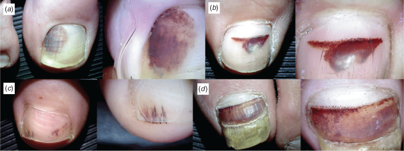
The features of subungual haemorrhage observed are listed in Table 1. The most common pattern of blood spots was a homogenous blood pattern (97%), followed by a globular blood spot pattern (78%) and streaks (34%). Most presentations (44; 75%) demonstrated a combination of blood patterns. A common combination of blood pattern specific to subungual haemorrhage includes distal streaks often associated with proximal clods or convex morphology. Multiple colours were recorded in the majority (37 cases; 63%) consistent with other reports of the dermoscopy of subungual haemorrhage.3 Forty-three (73%) had a clear proximal margin of normal nail bed and none had a longitudinal band extending from the proximal to distal nail (Figure 1).

|

|
None of the cases of subungual haemorrhage had any malignant dermoscopic characteristics associated with subungual melanoma. Thirty-two (54%) had nail dystrophy. Nail dystrophy can present as loss of nail translucency, discolouration or nail deformity (eg a thickened or cracked nail). Multiple repetitive trauma can by identified on dermoscopy by eliciting multiple partial horizontal lines, which can be suggestive of an underlying fracture of the nail plate. Although a feature of subungual melanoma, nail dystrophy has multifactorial causes of traumatic or inflammatory origin.
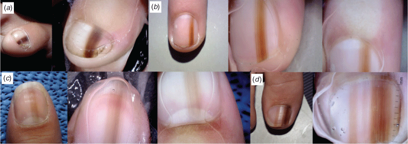
All melanocytic nail lesions displayed a pigmented longitudinal band extending from the proximal nail matrix to the distal nail plate. This was seen in benign melanonychia and in one of the two subungual melanomas (Figure 2). The other case of subungual melanoma enveloped the entire nail unit. Twenty-five cases of melanonychia covered less than one-third of the nail surface area. The most common colour was brown in 25 cases (68%). Twenty-six (70%) exhibited regular parallelism. Both cases of subungual melanoma displayed the micro-Hutchinson sign. There were no features of subungual haemorrhage, such as blood spots, streaks or peripheral fading.
Any doubtful lesions were monitored at 3- to 6-monthly intervals for reassurance.
Discussion
A structured approach is necessary to exclude malignancy and make a safe clinical diagnosis of subungual haemorrhage. Subungual haemorrhage can also be present as a feature of subungual melanoma.5
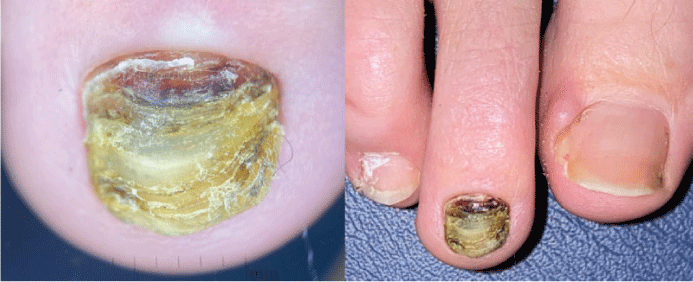
We can look at the assessment of subungual haemorrhage as a two-stage process. First, to identify the positive characteristics of subungual haemorrhage and second, to recognise the absence of malignant features. Apart from discolouration of the nail, irregular horizontal spots, lines or grooves across a portion of the nail plate are indicative of single or multiple traumas and hyperkeratosis of the tip of the digit implies callus formation. Haemorrhage affecting the second toenail is usually seen in people whose second toe is longer than the large toe and thus more prone to injury (Figure 3).
Subungual haemorrhage rarely presents as a longitudinal band, unlike melanonychia.7 It mostly presents with a homogenous blood pattern or globular blood spot pattern of red, purple, brown or black discolouration owing to an unstructured pattern of blood pooling beneath the nail plate.3 The variation of colour observed is related to the duration and the stage of healing.2 Pigmentation involving the entire nail or which does not follow this pattern presents diagnostic uncertainty.
There are no melanocytes in the nail bed. The physiology of melanin production within the proximal nail matrix gives rise to a longitudinal brown–black band within the nail plate, starting proximally and extending distally until the band covers the entire length of the nail plate.2,5 A clear proximal margin in the nail plate due to normal nail growth excludes a melanocytic lesion.
Dermoscopic features of nail unit melanoma include longitudinal bands with lines within the band that are irregular in colour, spacing and thickness.1–7 Other malignant dermoscopic features include a proximally wider triangular band indicating rapid growth, ulceration and micro-Hutchinson sign, with the latter reported as the most sensitive sign for subungual melanoma.2,5 These features are absent in subungual haemorrhage. Longitudinal nail pigmentation that affects one digit or with an onset that occurs in adulthood should be considered suspicious in the absence of other causes.8 In cases of persistent or non-resolving subungual haemorrhage, the possibility of nail bed tumours such as squamous cell carcinoma should be considered.
Our study confirms that combining macroscopic and dermoscopic characteristics is valuable in diagnosing subungual haemorrhage without the need for histological analysis. A pigmented nail lesion should always be evaluated for malignant melanocytic features.
The main limitation of our study is the small size of the cohort, which only included cases that were referred for a specialist opinion. It is very likely that most patients with subungual haemorrhage do not seek medical attention.
Competing interests
Dr Phillips has no conflicts of interest to declare. Dr Oakley is a contractor to MoleMap for private patients. MoleMap has been the provider of many images to the teledermatology service.
Funding
This research did not receive any specific funding.
Acknowledgements
The authors wish to acknowledge MoleMap New Zealand, provider of many images for the hospital’s teledermatology service.
References
[1] Nevares-Pomales OW, Sarriera-Lazaro C, Barrera-Llaurador J, et al. Pigmented lesions of the nail unit. Am J Dermatopathol. 2018; 40 793–804.| Pigmented lesions of the nail unit.Crossref | GoogleScholarGoogle Scholar | 30339563PubMed |
[2] Alessandrini A, Starace M, Piraccini B. Dermoscopy in the evaluation of nail disorders. Skin Appendage Disord. 2017; 3 70–82.
| Dermoscopy in the evaluation of nail disorders.Crossref | GoogleScholarGoogle Scholar | 28560217PubMed |
[3] Mun JH, Kim G, Jwa S, et al. Dermoscopy of subungual haemorrhage: its usefulness in differential diagnosis from nail-unit melanoma. Br J Dermatol. 2013; 168 1224–9.
| Dermoscopy of subungual haemorrhage: its usefulness in differential diagnosis from nail-unit melanoma.Crossref | GoogleScholarGoogle Scholar | 23302009PubMed |
[4] Koga H, Saida T, Uhara H. Key point in dermoscopic differentiation between early nail apparatus melanoma and benign longitudinal melanonychia. J Dermatol. 2011; 38 45–52.
| Key point in dermoscopic differentiation between early nail apparatus melanoma and benign longitudinal melanonychia.Crossref | GoogleScholarGoogle Scholar | 21175755PubMed |
[5] Phan A, Dalle S, Touzet S, et al. Dermoscopic features of acral lentiginous melanoma in a large series of 110 cases in a white population. Br J Dermatol. 2009; 162 765–71.
| Dermoscopic features of acral lentiginous melanoma in a large series of 110 cases in a white population.Crossref | GoogleScholarGoogle Scholar | 19922528PubMed |
[6] Benati E, Ribero S, Longo C, et al. Clinical and dermoscopic clues to differentiate pigmented nail bands: an International Dermoscopy Society study. J Eur Acad Dermatol Venereol. 2016; 31 732–6.
| Clinical and dermoscopic clues to differentiate pigmented nail bands: an International Dermoscopy Society study.Crossref | GoogleScholarGoogle Scholar | 27696528PubMed |
[7] Fountain JA. Recognition of subungual hematoma as an imitator of subungual melanoma. J Am Acad Dermatol. 1990; 23 773–4.
| Recognition of subungual hematoma as an imitator of subungual melanoma.Crossref | GoogleScholarGoogle Scholar | 2229520PubMed |
[8] Thomas L, Dalle S. Dermoscopy provides useful information for the management of melanonychia striata. Dermatol Ther. 2007; 20 3–10.
| Dermoscopy provides useful information for the management of melanonychia striata.Crossref | GoogleScholarGoogle Scholar | 17403255PubMed |


