Rural point-of-care ultrasound of the kidney and bladder: quality and effect on patient management
Garry Nixon 1 , Katharina Blattner 1 , Jill Muirhead 1 , Ngaire Kerse 21 Dunedin School of Medicine, University of Otago Dunedin, New Zealand
2 School of Population Health, University of Auckland, Tamaki, Auckland, New Zealand
Correspondence to: Gary Nixon, Dunedin School of Medicine, University of Otago Dunedin, New Zealand. Email: garry.nixon@otago.ac.nz
Journal of Primary Health Care 10(4) 324-330 https://doi.org/10.1071/HC18034
Published: 7 December 2018
Journal Compilation © Royal New Zealand College of General Practitioners 2018.
This is an open access article licensed under a Creative Commons Attribution-NonCommercial-NoDerivatives 4.0 International License.
Abstract
INTRODUCTION: Point-of-care ultrasound (POCUS) of the kidney and bladder are among the most commonly performed POCUS scans in rural New Zealand (NZ).
AIM: To determine the quality, safety and effect on patient care of POCUS of the kidney and bladder in rural NZ.
METHODS: Overall, 28 doctors in six NZ rural hospitals completed a questionnaire both before and after undertaking a POCUS scan over a 9-month period. The clinical records and saved ultrasound images were reviewed by a specialist panel.
RESULTS: The 28 participating doctors undertook 138 kidney and 60 bladder scans during the study. POCUS of the bladder as a test for urinary retention had a sensitivity of 100% (95% CI 88–100) and specificity of 100% (95% CI 93–100). POCUS of the kidney as a test for hydronephrosis had a sensitivity 90% (95% CI 74–96) and specificity of 96% (95% CI 89–98). The accuracy of other findings such as renal stones and bladder clot was lower. POCUS of the bladder appeared to have made a positive contribution to patient care in 92% of cases without evidence of harm. POCUS of the kidney benefited 93% of cases, although in three cases (2%), it may have had a negative effect on patient care.
DISCUSSION: POCUS as a test for urinary retention and hydronephrosis in the hands of rural doctors was technically straightforward, improved diagnostic certainty, increased discharges and overall had a positive effect on patient care.
KEYWORDS: POCUS; scans; hydronephrosis
| WHAT GAP THIS FILLS |
| What is already known: Ultrasound of the bladder and kidney are among the most commonly performed ultrasound examinations by rural NZ doctors. The safety and utility of ultrasound of the bladder and kidney has been proven in urban emergency departments. |
| What this study adds: Point-of-care ultrasound of the bladder for urinary retention, and of the kidney for hydronephrosis, are sensitive and specific tests in the rural setting. Rural point-of-care ultrasound of the bladder and kidney have a positive effect on patient care, which includes altering when to transfer patients to distant urban hospitals. |
Introduction
Point-of-care ultrasound (POCUS) of the kidney and bladder are the fourth and sixth most commonly performed POCUS examinations by New Zealand rural doctors respectively.1
POCUS of the kidney is also commonly performed in emergency departments, a practise that is supported by the emergency medicine literature.2 The principal finding being sought is hydronephrosis, usually when the differential diagnosis includes renal colic. POCUS of the bladder, as a test for urinary retention, is an even more straightforward examination, frequently performed by nursing and medical staff outside the radiology department.3
Those who practice POCUS in rural New Zealand consider it a valuable additional skill.1 This is principally because alternative diagnostic imaging is limited in NZ’s rural hospitals. Plain x-ray is often available only during normal working hours, few rural hospitals have formal ultrasound (performed by a trained sonographer and reported by a radiologist) and even fewer have immediate access to computed tomography.4
POCUS can be a technically difficult skill to learn. This is reflected in the formal training and accreditation processes that have been adopted by emergency medicine colleges around the world.5,6 POCUS is also resource-intensive, both with respect to equipment and training costs.
Few articles have been published on POCUS in the rural setting and we were unable to find any studies that evaluated the benefits (or otherwise) of POCUS to patient care in the rural context. The aim of this study is to evaluate POCUS of the kidney and bladder, in particular the ability of rural doctors to obtain and correctly interpret ultrasound images, the accuracy of their POCUS findings and the effect on diagnostic decision-making and patient management.
Methods
This study is a subgroup analysis of a larger study examining the safety and effect of POCUS on patient care in rural New Zealand.1,7
Twenty-eight rural generalist doctors, working in six rural NZ hospitals were enrolled in the study over a 9-month period in 2012. Three of the study hospitals were in the North Island and three in the South Island. The characteristics of the participating doctors (including their POCUS training) and the study hospitals, along with detailed methods are reported elsewhere.1
The participating doctors completed a questionnaire each time they used POCUS as part of their routine clinical duties. They completed the first section of the questionnaire prior to doing the POCUS examination and the second section after the POCUS (post-test). Both sections recorded: (1) the participating doctor’s estimation of the likelihood of the major diagnoses being considered (diagnostic probability); and (2) their planned disposition for the patient (i.e. discharge, admission to the local rural hospital or transfer to specialist base hospital by road or air). The differences between pre-test and post-test recordings were used to measure the effect of POCUS on diagnostic decision-making and patient disposition. The questionnaire also included the participating doctor’s impression of the image quality (self-reported scan quality) and their interpretation of the images (POCUS findings).
The investigators reviewed the clinical records of all cases in the 3 months following the study period. Where possible, definitive findings were determined based on the results of formal diagnostic imaging, the final diagnosis or a review of the saved POCUS images. Using the information in the clinical record, the investigators categorised the effect the POCUS had on patient management as either nil, some, significant, major or negative. ‘Some’ effect on patient management included confirming a diagnosis that was likely to have been made without the scan or ruling out an important, but very unlikely diagnosis. Examples of ‘significant’ effects on management included changing the intended patient disposition (e.g. deciding to discharge a patient that might have otherwise been admitted for observation) or leading to a diagnosis that was unclear prior to the scan. To meet the threshold for ‘major’ effect, there had to be evidence that the POCUS avoided major disability or death. ‘Negative’ effect was any situation in which it appeared the patient would have been better not to have had the POCUS scan; that is, it delayed the correct diagnosis or resulted in inappropriate clinical management. A specialist panel (comprising an emergency physician with an interest in POCUS, a radiologist and a sonographer) undertook a second review of the clinical record for selected cases. The panel reviewed all cases where investigators judged the effect to have been negative and any cases where the investigators were uncertain about the definitive findings or the effect POCUS had on patient care.
When they were available, the recorded POCUS images were reviewed by the sonographer on the specialist panel. The quality of the images was assessed (assessed scan quality) and the sonographer’s interpretation of the images was compared with the participant’s interpretation (scan interpretation).
Ethics approval was obtained from the NZ Multi Region Ethics Committee MEC/10/09/091.
Statistical analysis was undertaken using SPSS Version 23 (SPSS Inc., Chicago, IL, USA). Descriptive statistics were used to describe outcomes. True and false positive rates were derived by comparing participants’ POCUS findings and the definitive findings (gold standard). Sensitivities, specificities and positive and negative likelihood ratios were calculated using MEDCALC (MedCalc Software, Ostend, Belgium).8 Spearman correlation coefficient was used to establish the correlation between the patients’ pre- and post-scan disposition and between the post scan and actual disposition.
Results
The participating doctors undertook 138 kidney and 80 bladder scans over the study period. Electronic records of ultrasound images or clips were available for 124 kidney and 73 bladder scans.
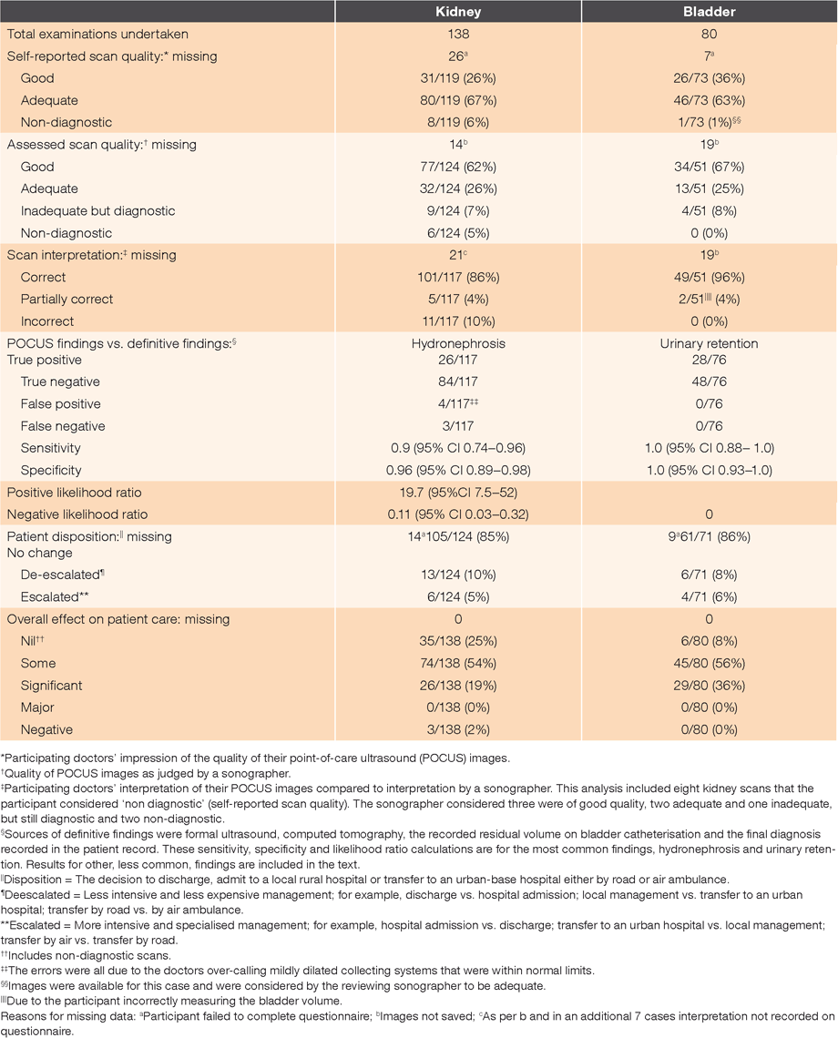
The results for both kidney and bladder scans are presented in Table 1. The reasons for missing data are detailed in the footnotes to Table 1. On most occasions, this was because participants did not complete parts of the questionnaire or record images.

|
The sonographer on the specialist panel was more likely than the participants themselves to consider the image quality for kidney scans to be ‘good’ (62% vs. 26% respectively). Both the sonographer and the participants considered a similar proportion of the kidney scans to be non-diagnostic (5% and 6% respectively) (Table 1). Similar results were obtained for bladder scans.
It was possible to compare the 139 POCUS kidney findings with definitive findings obtained from the clinical records. The calculated sensitivity, specificity and likelihood ratios for hydronephrosis, the most common finding being sought by participants (117/139 findings), is included in Table 1. On four occasions, the finding was a renal cyst. Three of these proved to be correct (true positive) but one was a false positive. On four occasions, participants concluded there was a stone in either the renal pelvis or the ureter. Only two of these were true positives; that is, for the remaining two cases, no stone was noted in the definitive findings. One renal mass was correctly identified.
The accuracy of the most common bladder finding, urinary retention, is presented in Table 1. On two occasions, participants reported finding blood clot in the bladder. On one occasion, they were correct (true positive), but on the other, they were incorrect (false positive).
Diagnostic probability
POCUS altered the probability of the principal diagnoses being considered for 97% of kidney cases and 86% of bladder cases. The overall effect on diagnostic probability is illustrated in Figures 1 and 2. Having undertaken POCUS, the participating doctors were more likely to be confident that the diagnosis being considered was present or absent (high or low probability). POCUS of the bladder was more likely than POCUS of the kidney to result in diagnostic certainty (post-test probability of 0% or 100%).

|

|
Patient disposition
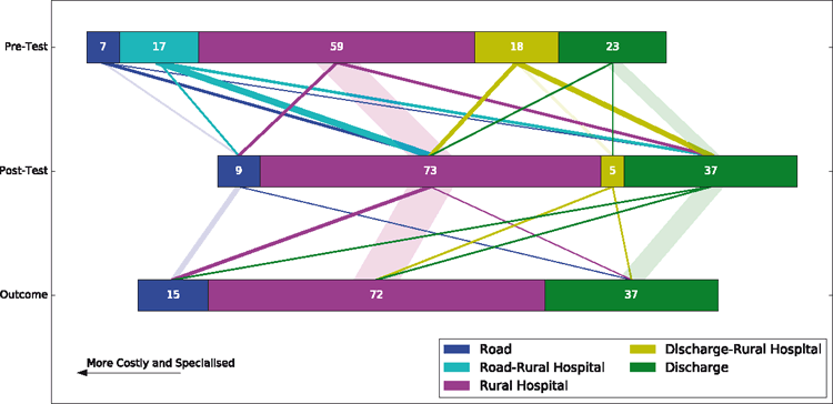
The effect of POCUS on the planned patient disposition and the actual disposition are illustrated in Figure 3 for kidney scans and Figure 4 for bladder scans.

|
There was a moderate correlation between pre-test and post-test disposition (Spearman correlation = 0.5, n = 124, P < 0.01) but a strong correlation between post-test and actual disposition (Spearman = 0.79, n = 124, P < 0.01) (Figure 3). The correlation between pre-test and actual disposition was the weakest 0.49, n= 124, P < 0.01.
There was a strong correlation between pre-test and post-test disposition (Spearman correlation = 0.62, n = 71, P < 0.01), but very strong correlation between post-test and actual disposition (Spearman = 0.98, n = 71, P < 0.01) (Figure 4). The correlation between pre- and actual disposition was 0.67, n = 71, P < 0.01.
The overall effect on patient care is presented in Table 1. On most occasions, POCUS was judged to have benefited patient care to at least some degree (73% of kidney scans and 92% of bladder scans). Three cases were identified where POCUS of the kidney may have negatively affected patient care. On one occasion, this was the result of missed hydronephrosis. The two other cases were due to missed stones. On no occasion did the specialist panel find definitive evidence of patient harm.
Discussion
This is the first study the authors are aware of that evaluates POCUS of the bladder and kidney in the rural context.
Bladder
In this study, POCUS proved to be a highly accurate test for urinary retention (sensitivity and specificity = 100%). This is considerably better than routine physical examination, which has a sensitivity of 80% and specificity of 50%.9 When taken together, these results explain why POCUS frequently increased diagnostic certainty and had a positive effect on patient care without evidence of harm. The low specificity of physical examination supports the authors’ impression that a considerable number of patients undergo urinary catheterisation that can be avoided when POCUS is available.
Obtaining and interpreting POCUS images of the bladder for urinary retention was technically straightforward. The only errors identified were with respect to measuring the bladder volume. On two occasions, the participants measured bladder depth in the transverse rather than the longitudinal plane. In the transverse plane, it is not possible to be sure that the depth is being measured perpendicular to the long axis, which can result in an inaccurate calculated volume.10 The correct technique for measuring bladder volume should be emphasised when teaching rural doctors POCUS skills.
Kidney
When used as a test for hydronephrosis, POCUS proved to be specific (96%) and reasonably sensitive (90%). This sensitivity and specificity are at the upper end of what has been reported in earlier studies on POCUS for hydronephrosis in the emergency medicine literature.2 The most common error, responsible for all four of the false positives, occurred when participants incorrectly interpreted mild dilatation of the collecting system (that was within normal limits) as hydronephrosis. This is particularly likely to occur when POCUS is performed on a patient with a full bladder. The importance of not overcalling mild dilatation of the collecting system and adequately preparing the patient should be reinforced by those teaching rural POCUS.
Hydronephrosis and urinary retention are the two urinary tract POCUS findings routinely taught to rural and emergency medicine doctors. Although numbers are too low to draw firm conclusions, the accuracy of POCUS was poor when the participants sought additional findings such as renal stones or blood clot in the bladder. This study does not provide evidence for the safe practise of rural POCUS of the renal tract beyond the findings of urinary retention and hydronephrosis.
Urinary retention and hydronephrosis are not in themselves findings that would mandate transferring a patient to a base hospital; renal colic and urinary retention can often be managed in the rural context. This is in contrast to other POCUS findings such as abdominal aortic aneurysm or free abdominal fluid following blunt abdominal trauma. We therefore did not expect to identify cases in which POCUS had a ‘major’ effect on patient care. POCUS examinations of the kidney and bladder did, however, alter planned patient disposition more often than might have been expected (15 and 14% respectively). As shown in Figures 3 and 4, this included both escalating and de-escalating the level of patient care. Overall, POCUS increased the number of patients who were discharged and saw a similar number of patients transferred to a base hospital; suggesting a reduction in health service costs.
In this study, POCUS proved to be a useful diagnostic test for patients who may have urinary retention or hydronephrosis in the rural context.
COMPETING INTERESTS
None.
ACKNOWLEDGEMENTS
The authors wish to thank Dr Rosylne McKechnie and Ms Bron Hunt for administering the data collection and analysis, and for liaising with the participants.
References
[1] Nixon G, Blattner K, Lawrenson R, Kerse N. The scope of point-of-care ultrasound practice in rural New Zealand. J Prim Health Care. 2018; 10 224–36.| The scope of point-of-care ultrasound practice in rural New Zealand.Crossref | GoogleScholarGoogle Scholar |
[2] Dalziel PJ, Noble VE. Bedside ultrasound and the assessment of renal colic: a review. Emerg Med J. 2013; 30 3–8.
| Bedside ultrasound and the assessment of renal colic: a review.Crossref | GoogleScholarGoogle Scholar |
[3] Davis C, Chrisman J, Walden P. To scan or not to scan? Detecting urinary retention. Nursing Made Incredibly Easy. 2012; 10 53–4.
| To scan or not to scan? Detecting urinary retention.Crossref | GoogleScholarGoogle Scholar |
[4] Nixon G, Samaranayaka A, de Graaf B, McKechnie R, Blattner K, Dovey S. Geographic disparities in the utilisation of computed tomography scanning services in southern New Zealand. Health Policy. 2014; 118 222–8.
| Geographic disparities in the utilisation of computed tomography scanning services in southern New Zealand.Crossref | GoogleScholarGoogle Scholar |
[5] Council of Australasian College for Emergency Medicine. Policy on the focused use of focused ultrasound in emergency medicine. Melbourne: Australasian College for Emergency Medicine; 2016.
[6] American College of Emergency Physicians Ultrasound Guidelines: Emergency, Point-of-Care and clinical ultrasound guidelines in medicine. Ann Emerg Med. 2017; 69 e27–e54.
[7] Nixon G, Blatter K, Koroheke-Rogers M,, et al. Point-of-care ultrasound in rural New Zealand: safety, quality and impact on patient management. Aust J Rural Health. 2018; 26 342–9.
| Point-of-care ultrasound in rural New Zealand: safety, quality and impact on patient management.Crossref | GoogleScholarGoogle Scholar |
[8] MEDCALC®. 2018 Easy-to-use statistical software. Ostend: Belgium; 2018 [cited 2018 October 26]. Available from: https://www.medcalc.org/calc/diagnostic_test.php
[9] Weatherall M, Harwood M. The accuracy of clinical assessment of bladder volume. Arch Phys Med Rehabil. 2002; 83 1300–2.
| The accuracy of clinical assessment of bladder volume.Crossref | GoogleScholarGoogle Scholar |
[10] Hvarness H, Skjoldbye B, Jakobsen H. Urinary bladder volume measurements: comparison of three ultrasound calculation methods. Scand J Urol Nephrol. 2002; 36 177–81.
| Urinary bladder volume measurements: comparison of three ultrasound calculation methods.Crossref | GoogleScholarGoogle Scholar |


