Michael Desmond Connole: Veterinary Mycologist
Justine S GibsonThe University of Queensland, School of Veterinary Science, Gatton Qld 4343, Australia
Tel: +61 7 5460 1830
Fax: +61 7 5460 1922
Email: gibson.j@uq.edu.au
Microbiology Australia 34(1) 47-50 https://doi.org/10.1071/MA13015
Published: 20 March 2013
Veterinary mycology is often a neglected field in veterinary medicine with many veterinarians treating infections empirically, or failing to send samples to diagnostic laboratories for identification. Few researchers undertake projects in this field. However, between 1968 and 1992 there was a major veterinary mycology laboratory in Australia. This laboratory was established at the Animal Research Institute, Yeerongpilly by Michael Desmond Connole known to friends and colleagues as “Des”. Des was actively engaged in research, training, diagnosis and control of veterinary mycoses in Australia for nearly 50 years. He is an international figure in the field of mycoses and has over 45 publications including: “A review of animal mycosis in Australia”, which was published in Mycopathologia in 1990. Des specialised in the isolation of fungi including dermatophytes, zygomycetes, pathogenic hyphomycetes and mycotoxin producers. This article honours Des’s contribution to veterinary mycology in Australia and internationally.
Des had humble beginnings growing up in Camp Hill in Brisbane. He completed his junior pass at St Laurence’s in South Brisbane and then entered the public service as a clerk in 1943. He completed his senior pass by studying nights and Saturday mornings at the Queensland Teacher’s Training College in Edward Street, Brisbane.
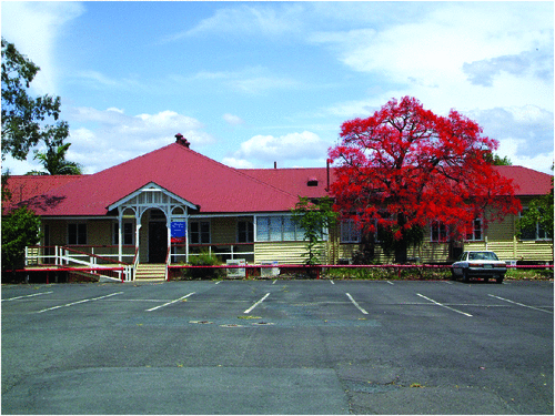
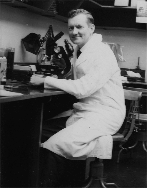
In 1947, Des started working as a cadet science graduate (bacteriologist) at the Animal Research Institute (ARI) at Yeerongpilly (Figure 1) where he was supervised by Geoffrey Clive Simmons who was the chief bacteriologist and first graduate bacteriologist to be employed at ARI. In 1957, Des graduated from The University of Queensland with a Bachelor of Science, majoring in Bacteriology (Figure 2). Des worked as a diagnostic bacteriologist, from 1957–1963, identifying the causal agents (bacteria, viruses, protozoa and fungi) of animal diseases. During his spare time he was told to “work on mycology as there has been no work done on veterinary mycology”. Thus, Des’s great love of veterinary mycology began.

|

|
Des’s first solo publication detailed a natural ringworm infection in the guinea pig colony at the ARI due to Trichophyton mentagrophytes. The paper describes the progression of lesions, and the diagnosis of the dermatophytes via microscopic examination and culture of skin scrapings and hair1. Much of Des’s work involved the study of dermatophytes. In 1959, he identified Trichophyton verrucosum from a bovine skin scraping. This was the first time it had been isolated from cattle in Australia and the isolate was sent it to the London School of Hygiene and Tropical Medicine, to confirm his identification. Previously, T. verrucosum had only been isolated from human cases in Victoria, Australia. Humans with this dermatophyte infection worked on farms and with animals. Hence, the isolation of the agent from cattle was a major breakthrough and identified a zoonotic link. This led to a survey of bovine ringworm (1960–1962) where all field staff submitted suspected ringworm cases from bovines. Des was able to identify 32 strains of T. verrucosum from field specimens and 14 strains from stock at the institute. He identified that T. verrucosum was the usual agent of cattle ringworm and occurred in all parts of Queensland, in both dairy and beef cattle and while it could occur in all age groups, young cattle were most at risk2.
In 1963, Des went on study leave to the University of Glasgow where he worked under Professor J. C. Gentles. Here he developed his skills in diagnostic mycology, teaching and research (Figure 3). During this period he studied dermatophytes and keratinophilic fungi of dogs and cats. This work led to two publications, the first describing the “hairbrush sampling” technique which he performed on 154 animals to detect the presence of keratinophilic fungi3 and the second was published with Christine Dawson who continued his work, with some variations in culture methods and examined another series of animals at approximately the same time the following year4. The “hairbrush sampling” technique was identified as adequate if the fungus is present in quantity, as typically occurs in dermatophyte infections. On his way back to Australia from Glasgow, Des spent seven weeks at the Central Veterinary Laboratory, Weybridge, England, and visited other laboratories, universities and industries in the United Kingdom, visited the Pasteur institute, Paris and laboratories in the United States of America including the Centre for Disease Control, Atlanta, Georgia.

|
On returning to Australia, Des published data from a survey of dermatophytes from dogs and cats. He was able to identify Microsporum canis and M. gypseum. This was the first record of M. gypseum infection in dogs in Australia2. In 1968, Des published the first record of a T. mentagrophytes infection in a dog in Australia5.
In 1964, Des moved to Townsville where he worked as the diagnostic bacteriologist and mycologist at the Oonoonba Animal Health Station, until 1968. Here he developed expertise in identifying potentially toxic fungi (esp. Aspergillus) presented in animal feedstuffs6.
In 1967, via Invitation of the Commonwealth Bureau of Animal Health, Des was asked to perform the first Australian review of animal mycoses7. At this time, mycotic diseases of animals were of economic importance and some as zoonoses were of public health interest. There was no satisfactory treatment for many mycotic infections and the epidemiology of mycoses was virtually unknown. Des with co-author L.A.Y. Johnston reviewed veterinary superficial and systemic mycoses, and mycotoxicoses from Australia7.
In 1968, Des returned to ARI, as the Senior Bacteriologist (Mycology). In the mycology unit at ARI he diagnosed dermatophytes, hyphomycetes, zygomycetes, and examined feedstuffs for toxigenic fungi. He was sent fungal cultures from intrastate, interstate and overseas.
Throughout his career Des studied dermatophytosis in horses. Ringworm in horses is undesirable as horses with detectable infections are not allowed to race and infections are potentially transmissible to humans. Des was involved in the first isolation of T. equinum the usual ringworm agent affecting horses in Queensland in 1960–19632, but it was not until 1984 after an outbreak of severe ringworm in horses in the Oakey area, that this was recognised as a Trichophyton equinum var. equinum infection8,9. Des was also the first to isolate Microsporum gypseum from a skin scraping from a four year old pony from Central Queensland in 196610. In 1974, during an outbreak of M. gypseum infections, Des with equine veterinarian Dr Reginald “Reg” Pascoe described a number of natural and experimental infections in horses11. This body of work described gross lesions, diagnosis, microscopic and cultural characteristics of dermatophytes, and the epidemiology of infection including risk factors such as biting insects, and moist atmospheric conditions. They were able to implicate fomites like girth straps and saddle cloths in the transmission of infections11.
Des also isolated M. nanum from pigs in Queensland and investigated experimental transmission of the dermatophyte12. Des performed a number of studies examining fungal metabolites to use as insecticides against the cattle tick (Boophilus microplus)13 and the sheep blowfly (Lucilia cuprina)14,15. He also continued to investigate mycotoxins, and reported on a number of cases involving aflatoxicosis and isolation of mycotoxins in animal feeds16–19.
Des also identified a number of rare opportunistic mycoses such as a Drechslera rostrata infection in a cow, which was first infection of this type recorded in Australia20, and reported on mycotic nasal granumalomas in cattle from 1966–197521. He identified Conidiobolus incrongruus as the cause of rhinocerbral and nasal granulomas in sheep22. He reported on equine phycomycosis23 and identified Moniliella suaveolens as the cause of opportunistic granulomas in cats24, which had not been previously identified from animals or man. He even helped identify green algae from green lymph nodes from cattle with lymphadenitis25.
Des was the supervising bacteriologist at the ARI during 1974–1982 and continued his diagnostic work and also presented courses to medical mycology students, postgraduate students and trained veterinary mycologists from Australia and overseas. In 1982, on behalf of the Australian Development Assistance Bureau, Canberra, Des carried out a short-term consultancy in the Mycology Department of the Animal Disease Institute, Bogor, Indonesia. He spent time training technicians in mycology skills, in what was quite a challenging tropical climate, with many contamination problems, including fungi growing on microscope lenses.
In 1990, he was asked to perform a second review on animal mycoses in Australia. This review26 covered literature on mycoses in animals in Australia since the last review published in 1967.
In addition to his outstanding work as a diagnostic bacteriologist, mycologist and researcher, Des played an important role in the Australian Society of Microbiology. He was a founding member and first Treasurer of the Queensland branch. In 1978, he became a foundation member of the Australasian Federation of Medical and Veterinary Mycology a special interest group of the ASM. He served on various committees and contributed to many conferences. In 1982, he received the first Churchill fellowship awarded in Australia in the field of medical and veterinary mycology and travelled to New Zealand to study developments in animal and human mycology. Des has also played an active role with the International Society for Human and Animal Mycology (ISHAM). He has attended 13 ISHAM congresses since 1964, including this year’s meeting in Berlin. Des retired in 1992, after nearly 50 years of public service. He continued to work for a number of years delivering lectures, consulting and refereeing journal articles. Now he enjoys his retirement with his wife, Kathy, though he still regularly attends mycology conferences.
Acknowledgements
I acknowledge Des and his wife Kath for their assistance and support in compiling this article.
References
[1] Connole, M.D. (1963) Ringworm in guinea pigs due to Trichophyton mentagrophytes. Qld J. Argic. Sci. 20, 293–297.[2] Connole, M.D. (1963) A review of dermatomycoses of animals in Australia. Aust. Vet. J. 39, 130–134.
| A review of dermatomycoses of animals in Australia.Crossref | GoogleScholarGoogle Scholar |
[3] Connole, M.D. (1965) Keratinophilic fungi on cats and dogs. Sabouraudia 4, 45–48.
| Keratinophilic fungi on cats and dogs.Crossref | GoogleScholarGoogle Scholar | 1:STN:280:DyaF2s%2FosFWjsw%3D%3D&md5=a80f922645a4fda0a6ad997b87157814CAS |
[4] Gentles, J.C. et al. (1966) Keratinophilic fungi on cats and dogs. II. Sabouraudia 4, 171–175.
| Keratinophilic fungi on cats and dogs. II.Crossref | GoogleScholarGoogle Scholar |
[5] Connole, M.D. (1968) Ringworm due to Trichophyton mentagrophytes in a dog. Aust. Vet. J. 44, 528.
| Ringworm due to Trichophyton mentagrophytes in a dog.Crossref | GoogleScholarGoogle Scholar | 1:STN:280:DyaF1M%2FlsF2hsw%3D%3D&md5=ce9a88b17e27c86c9594a3fb6cfb925dCAS |
[6] Connole, M.D. and Hill, M.W. (1970) Aspergillus flavus contaminated sorghum grain as a possible cause of aflatoxicosis in pigs. Aust. Vet. J. 46, 503–505.
| Aspergillus flavus contaminated sorghum grain as a possible cause of aflatoxicosis in pigs.Crossref | GoogleScholarGoogle Scholar | 1:STN:280:DyaE3M%2FjslOmsQ%3D%3D&md5=2ad96a74ccf1b78874b49d806b8e73cfCAS |
[7] Connole, M.D. and Johnston, L.A.Y. (1967) A review of animal mycoses. Vet. Bull. 37, 145–153.
[8] Smith, J.M. et al. (1968) Trichophyton equinum var. autotrophicum; its characteristics and geographical distribution. Sabouraudia 6, 296–304.
| Trichophyton equinum var. autotrophicum; its characteristics and geographical distribution.Crossref | GoogleScholarGoogle Scholar | 1:STN:280:DyaF1M%2Fmtlajtg%3D%3D&md5=7a93c1d6fd2ffb8d796aeb139631cb9eCAS |
[9] Connole, M.D. and Pascoe, R.R. (1984) Recognition of Trichophyton equinum var. equinum infection of horses. Aust. Vet. J. 61, 94.
| Recognition of Trichophyton equinum var. equinum infection of horses.Crossref | GoogleScholarGoogle Scholar | 1:STN:280:DyaL2c3ls1entg%3D%3D&md5=e7b119baef3a2ce4cc6b15c978046c16CAS |
[10] Connole, M.D. (1967) Microsproum gypseum ringworm in a horse. Aust. Vet. J. 43, 118.
| Microsproum gypseum ringworm in a horse.Crossref | GoogleScholarGoogle Scholar |
[11] Pascoe, R.R. and Connole, M.D. (1974) Dermatomycosis due to Microsporum gypseum in horses. Aust. Vet. J. 50, 380–383.
| Dermatomycosis due to Microsporum gypseum in horses.Crossref | GoogleScholarGoogle Scholar | 1:STN:280:DyaE2M%2Fnt1eltw%3D%3D&md5=83a557e833ae4587653f8d503bb9f83eCAS |
[12] Connole, M.D. and Baynes, I.D. (1966) Ringworm caused by Microsporum nanum in pigs in Queensland. Aust. Vet. J. 42, 19–24.
| Ringworm caused by Microsporum nanum in pigs in Queensland.Crossref | GoogleScholarGoogle Scholar | 1:STN:280:DyaF2s%2FhvFWlsw%3D%3D&md5=73ca38a4b98b04d9404ead817075826bCAS |
[13] Connole, M.D. (1969) Effect of fungal extracts on the cattle tick, Boophilus microplus. Aust. Vet. J. 45, 207.
| Effect of fungal extracts on the cattle tick, Boophilus microplus.Crossref | GoogleScholarGoogle Scholar | 1:STN:280:DyaF1M7osFyjtw%3D%3D&md5=e974ece6be6bc7f72221365a7a992653CAS |
[14] Blaney, B.J. et al. (1985) Fungal metabolites with insecticidal properties I. Relative toxicity of extracts of fungal cultures to sheep blowfly Lucilia cuprina IWeid. Gen. Appl. Entom. 17, 42–46.
[15] Green, P.E. et al. (1989) Identification and preliminary evaluation of viriditoxin, a metabolite of Paecilomyces varioti as an insecticide for sheep blowfly, Lucilia cuprina (Wied). Gen. Appl. Entom 21, 33–37.
[16] Connole, M.D. et al. (1981) Mycotoxins and mycotoxic fungi in Queensland stockfeeds 1971-80. Aust. Vet. J. 57, 314–318.
| Mycotoxins and mycotoxic fungi in Queensland stockfeeds 1971-80.Crossref | GoogleScholarGoogle Scholar | 1:STN:280:DyaL387otF2hsQ%3D%3D&md5=71b4438cf0693145ced8c1a428d20455CAS |
[17] McKenzie, R.A. et al. (1981) Acute aflatoxicosis in calves fed peanut hay. Aust. Vet. J. 57, 284–286.
| Acute aflatoxicosis in calves fed peanut hay.Crossref | GoogleScholarGoogle Scholar | 1:STN:280:DyaL38%2FptVymsA%3D%3D&md5=74ea530b074d8778192a559f88cfc8e4CAS |
[18] Blaney, B.J. et al. (1989) Aflatoxin and cyclopiazonic acid production by Queensland isolates of Aspergillus flavus and Aspergillus parasiticus. Aust. J. Agric. Res. 40, 395–400.
| Aflatoxin and cyclopiazonic acid production by Queensland isolates of Aspergillus flavus and Aspergillus parasiticus.Crossref | GoogleScholarGoogle Scholar | 1:CAS:528:DyaL1MXltFahtrc%3D&md5=61c803264d3d8724be317974439aca0bCAS |
[19] Ketterer, P.J. et al. (1975) Canine aflatoxicosis. Aust. Vet. J. 51, 355–357.
| Canine aflatoxicosis.Crossref | GoogleScholarGoogle Scholar | 1:STN:280:DyaE28%2FjvFaluw%3D%3D&md5=de04d46817ab9f6e2e1aec11f64a231bCAS |
[20] Pritchard, D. et al. (1977) Eumycotic mycetoma due to Drechslera rostrata infection in a cow. Aust. Vet. J. 53, 241–244.
| Eumycotic mycetoma due to Drechslera rostrata infection in a cow.Crossref | GoogleScholarGoogle Scholar | 1:STN:280:DyaE2s3ms1ehug%3D%3D&md5=87d6fe28e5a0e1fad21a5b09a3195f17CAS |
[21] McKenzie, R.A. and Connole, M.D. (1977) Mycotic nasal granuloma in cattle. Aust. Vet. J. 53, 268–270.
| Mycotic nasal granuloma in cattle.Crossref | GoogleScholarGoogle Scholar | 1:STN:280:DyaE2s3ms1eguw%3D%3D&md5=2ecb203dc131afe4c3bcfb0eb06a7250CAS |
[22] Ketterer, P.J. et al. (1992) Rhinocerebral and nasal zygomycosis in sheep caused by Conidiobolus incongruus. Aust. Vet. J. 69, 85–87.
| Rhinocerebral and nasal zygomycosis in sheep caused by Conidiobolus incongruus.Crossref | GoogleScholarGoogle Scholar | 1:STN:280:DyaK383pslSmtw%3D%3D&md5=c75c5778ddcc2e8293fbcce8bddc58edCAS |
[23] Connole, M.D. (1973) Equine phycomycosis. Aust. Vet. J. 49, 214–215.
| Equine phycomycosis.Crossref | GoogleScholarGoogle Scholar | 1:STN:280:DyaE3s7ps1Cmtw%3D%3D&md5=bd97d14af74d2f87817b466eec530830CAS |
[24] McKenzie, R.A. et al. (1984) Subcutaneous phaeohyphomycosis caused by Moniliella suaveolens in two cats. Vet. Pathol. 21, 582–586.
| 1:STN:280:DyaL2M%2Fpt1agtA%3D%3D&md5=b15304ac7e1e836ebb33210a6f5ee85bCAS |
[25] Rogers, R.J. et al. (1980) Lymphadenitis of cattle due to infection with green algae. J. Comp. Pathol. 90, 1–9.
| Lymphadenitis of cattle due to infection with green algae.Crossref | GoogleScholarGoogle Scholar | 1:STN:280:DyaL3c3ivValtw%3D%3D&md5=9c9629fb5a4f82f639829048f9b0f942CAS |
[26] Connole, M.D. (1990) Review of animal mycoses in Australia. Mycopathologia 111, 133–164.
| Review of animal mycoses in Australia.Crossref | GoogleScholarGoogle Scholar | 1:STN:280:DyaK3M%2Fkt12ntA%3D%3D&md5=5437652dbe488f54636a9f8d87d6d5deCAS |
Biography
Justine Gibson is a lecturer in Veterinary Bacteriology and Mycology at the School of Veterinary Science at The University of Queensland. Her research interests are in veterinary microbiology and molecular biology.


