

Neurological disease caused by flavivirus infections
Tristan Gibbs A B C and David J Speers A BA Department of Microbiology, PathWest Laboratory Medicine WA (LMWA), Queen Elizabeth II (QEII) Medical Centre, Hospital Avenue, Nedlands, WA 6009, Australia
B School of Medicine, Faculty of Health and Medical Sciences, University of Western Australia, 35 Stirling Highway, Crawley, WA 6009, Australia
C Email: tristan.gibbs@health.wa.gov.au
Microbiology Australia 39(2) 99-102 https://doi.org/10.1071/MA18029
Published: 6 April 2018
The Flavivirus genus contains dozens of species with varying geographical distributions. Most flavivirus infections in humans are asymptomatic or manifest as a non-specific febrile illness, sometimes accompanied by rash or arthralgia. Certain species are more commonly associated with neurological disease and may be termed neurotropic flaviviruses. Several flaviviruses endemic to Australia and our near northern neighbours are neurotropic, such as Murray Valley encephalitis virus, West Nile (Kunjin) virus and Japanese encephalitis virus. Flavivirus neurological disease ranges from self-limiting meningitis to fulminant encephalitis causing permanent debilitating neurological sequelae or death. The recent Zika virus outbreak in South America has highlighted the dramatic effects of flavivirus neurotropism on the developing brain. This article focuses on the neurotropic flaviviruses endemic to Australia and those of international significance.
Neurotropic flaviviruses of Australia
Murray Valley encephalitis virus (MVEV) and Kunjin virus (WNVKUN), a clade of West Nile virus (WNV), are endemic to Australia, causing sporadic neurological disease and occasionally outbreaks associated with increased mosquito activity during the wet season. Both viruses are maintained in mosquito-waterbird cycles primarily in northern Western Australia, the top end of the Northern Territory and possibly northern Queensland1,2. However, heavy rainfall with flooding may lead to spread of these viruses into normally arid areas, carried by waterbirds3. There were several outbreaks of MVEV on the east coast of Australia in 1951 and 1974 along the Murray-Darling River basin that gave the virus its name. The last widespread outbreak of MVEV occurred in 2011, with 17 cases across WA, NT, SA and NSW.
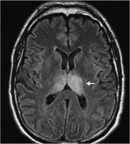
It is estimated that between 1 : 150 and 1 : 1000 of those infected with MVEV will develop encephalitis, which may manifest as seizures, altered mental state, focal neurological abnormalities, coma, or flaccid paralysis1. Characteristic thalamic involvement may be seen on brain MRI (Figure 1) but the changes may take several days to develop. The case fatality rate is 15–30% with long-term neurological sequelae occurring in 30–50%4. WNVKUN follows a similar epidemiological and clinical pattern to MVEV, although neurological disease tends to be milder, with no recorded cases of fatal infection3.
|
The public health management of MVEV and WNVKUN includes surveillance using antibody testing in sentinel chicken flocks and, more recently, detection of viral RNA in trapped mosquitoes. These data serve as an early warning system for increased flavivirus activity and potential increased risk of human cases to prompt timely public health advice to the public for mosquito avoidance measures and to medical services for appropriate diagnostic test ordering5.
Neurotropic flaviviruses outside Australia
The most important neurotropic flaviviruses for human health worldwide are Japanese encephalitis virus (JEV), WNV and Zika virus (ZIKV). Infection with these viruses should be suspected in travellers returning from areas where these viruses are endemic who develop compatible symptoms within the relevant incubation period. Depending on the travel and exposure history, the differential diagnosis may include other neurotropic arboviruses, such as alphaviruses and bunyaviruses.
JEV is widespread in Asia with over 50 000 cases of JEV encephalitis reported every year, mostly in children, with a case-fatality rate of 20–30% and neurological sequelae in 70% of survivors6. JEV vaccination programs have been implemented in several countries including China, India, Japan and Thailand, though data on the effectiveness of these programs is still emerging.
WNV was endemic only to Africa and Eurasia until it was introduced to New York in 1999, then spread across North and South America7. Since 2007, a yearly average of over 1000 cases of neurological disease has been reported in the USA8. Between 1 : 150 and 1 : 240 of those infected develop neurological disease9,10, with the risk increasing with age11.
ZIKV was restricted to Africa and Southeast Asia until outbreaks occurred in Micronesia in 2007, French Polynesia in 2013 and Brazil in early 2015. By July 2016, more than 150 000 cases were reported in Brazil and the virus had spread to central and South America and the Caribbean12,13. It is estimated that 80% of Zika infections are asymptomatic. An increase in cases of Guillain-Barré syndrome (GBS) was reported following the outbreak in French Polynesia and subsequently also reported in Brazil, Colombia, and El Salvador13,14. Less commonly, ZIKV may cause meningoencephalitis and transverse myelitis in adults.
During the 2015 outbreak in Brazil, an increase in congenital microcephaly was noted and suspected to be linked to maternal Zika virus infection. Retrospective investigation of the outbreak in French Polynesia outbreak also found an increase in microcephaly notifications. Further data support a causal link between maternal ZIKV infection and congenital Zika syndrome, manifestations of which include microcephaly, obstructive hydrocephalus, cerebral calcifications, congenital contractures, and hypertonia13,15–17. The full neurological spectrum of congenital Zika syndrome will become clearer with longer term follow-up studies of the ZIKV infected infants.
Dengue virus has the most extensive geographical distribution of all the flaviviruses known to infect humans and is the most frequently diagnosed flavivirus infection in travellers returning to Australia. While severe dengue infection usually manifests as shock from plasma leakage or haemorrhage, rare neurological complications of dengue such as encephalitis, meningitis, transverse myelitis and Guillain–Barré syndrome have also been described18.
See Table 1 for further information on selected neurotropic flaviviruses.

|
Laboratory diagnosis of flavivirus neurological disease
IgM antibody may be the earliest serological marker in flavivirus encephalitis21 and, in patients who develop an encephalitic illness, detection of flavivirus IgM antibody in serum confirms the diagnosis22. Recent flavivirus infection is usually confirmed by IgG or haemagglutination-inhibiting antibody seroconversion or a significant titre increase, or detection of flavivirus by cell culture or RT-PCR. Interpretation of flavivirus antibody titres is made more difficult in those with immunological memory from previous flavivirus exposure due to ‘original antigenic sin’. Given the high proportion of mild or asymptomatic cases, confirmation of infection with a neurotropic flavivirus does not necessarily indicate neurological disease. Where invasive specimens are available, definitive diagnosis can be made by detection of flavivirus in cerebrospinal fluid (CSF) or brain biopsy by cell culture or RT-PCR, or by detection of specific flavivirus IgM antibody in CSF.
When requesting diagnostic testing for flavivirus, it is important to consider the range of likely infecting flaviviruses, which is seasonally and geographically dependent. Relevant clinical and travel history should be provided to the testing laboratory to assist with test selection and interpretation.
References
[1] Selvey, L.A. et al. (2014) The changing epidemiology of Murray Valley encephalitis in Australia: the 2011 outbreak and a review of the literature. PLoS Negl. Trop. Dis. 8, e2656.| The changing epidemiology of Murray Valley encephalitis in Australia: the 2011 outbreak and a review of the literature.Crossref | GoogleScholarGoogle Scholar |
[2] Prow, N.A. (2013) The changing epidemiology of Kunjin virus in Australia. Int. J. Environ. Res. Public Health 10, 6255–6272.
| The changing epidemiology of Kunjin virus in Australia.Crossref | GoogleScholarGoogle Scholar |
[3] Smith, D.W. et al. (2011) The viruses of Australia and the risk to tourists. Travel Med. Infect. Dis. 9, 113–125.
| The viruses of Australia and the risk to tourists.Crossref | GoogleScholarGoogle Scholar |
[4] Knox, J. et al. (2012) Murray Valley encephalitis: a review of clinical features, diagnosis and treatment. Med. J. Aust. 196, 322–326.
| Murray Valley encephalitis: a review of clinical features, diagnosis and treatment.Crossref | GoogleScholarGoogle Scholar |
[5] Selvey, L.A. et al. (2014) Rainfall and sentinel chicken seroconversions predict human cases of Murray Valley encephalitis in the north of Western Australia. BMC Infect. Dis. 14, 672.
| Rainfall and sentinel chicken seroconversions predict human cases of Murray Valley encephalitis in the north of Western Australia.Crossref | GoogleScholarGoogle Scholar |
[6] Campbell, G.L. et al. (2011) Estimated global incidence of Japanese encephalitis: a systematic review. Bull. World Health Organ. 89, 766–774.
| Estimated global incidence of Japanese encephalitis: a systematic review.Crossref | GoogleScholarGoogle Scholar |
[7] Gould, E.A. and Solomon, T. (2008) Pathogenic flaviviruses. Lancet 371, 500–509.
| Pathogenic flaviviruses.Crossref | GoogleScholarGoogle Scholar | 1:CAS:528:DC%2BD1cXhslelur0%3D&md5=1e8a56f3345d83a5610baa9228609749CAS |
[8] Centers for Disease Control and Prevention (2018) West Nile virus statistics. https://www.cdc.gov/westnile/statsmaps/cumMapsData.html (accessed 29 January 2018).
[9] Williamson, P.C. et al. (2017) Incidence of West Nile virus infection in the Dallas–Fort Worth metropolitan area during the 2012 epidemic. Epidemiol. Infect. 145, 2536–2544.
| Incidence of West Nile virus infection in the Dallas–Fort Worth metropolitan area during the 2012 epidemic.Crossref | GoogleScholarGoogle Scholar | 1:CAS:528:DC%2BC2sXhsFWqu7jJ&md5=6fd8d01a1fce50db8e3ece53706b9aa0CAS |
[10] Petersen, L.R. et al. (2003) West Nile virus. JAMA 290, 524–528.
| West Nile virus.Crossref | GoogleScholarGoogle Scholar |
[11] Krow-Lucal, E. et al. (2017) West Nile virus and other nationally notifiable arboviral diseases – United States, 2015. MMWR Morb. Mortal. Wkly. Rep. 66, 51–55.
| West Nile virus and other nationally notifiable arboviral diseases – United States, 2015.Crossref | GoogleScholarGoogle Scholar |
[12] da Silva, I.R.F. et al. (2017) Neurologic complications associated with the Zika virus in Brazilian adults. JAMA Neurol. 74, 1190–1198.
| Neurologic complications associated with the Zika virus in Brazilian adults.Crossref | GoogleScholarGoogle Scholar |
[13] Musso, D. and Gubler, D.J. (2016) Zika Virus. Clin. Microbiol. Rev. 29, 487–524.
| Zika Virus.Crossref | GoogleScholarGoogle Scholar | 1:CAS:528:DC%2BC1cXmsVygsLg%3D&md5=18d0f4fabeaae21b74d53cf3f71ed0c4CAS |
[14] Parra, B. et al. (2016) Guillain-Barré syndrome associated with Zika virus infection in Colombia. N. Engl. J. Med. 375, 1513–1523.
| Guillain-Barré syndrome associated with Zika virus infection in Colombia.Crossref | GoogleScholarGoogle Scholar |
[15] Brasil, P. et al. (2016) Zika virus infection in pregnant woman in Rio de Janeiro. N. Engl. J. Med. 375, 2321–2334.
| Zika virus infection in pregnant woman in Rio de Janeiro.Crossref | GoogleScholarGoogle Scholar |
[16] Chimelli, L. and Avvad-Portari, E. (2018) Congenital Zika virus infection: a neuropathological review. Childs Nerv. Syst. 34, 95–99.
| Congenital Zika virus infection: a neuropathological review.Crossref | GoogleScholarGoogle Scholar | 1:STN:280:DC%2BC1M3kt1ejsg%3D%3D&md5=389a781a45c963a9565e80ff0fffed8bCAS |
[17] Moore, C.A. et al. (2017) Characterizing the pattern of anomalies in congenital Zika syndrome for pediatric clinicians. JAMA Pediatr. 171, 288–295.
| Characterizing the pattern of anomalies in congenital Zika syndrome for pediatric clinicians.Crossref | GoogleScholarGoogle Scholar |
[18] Carod-Artal, F.J. et al. (2013) Neurological complications of dengue virus infection. Lancet Neurol. 12, 906–919.
| Neurological complications of dengue virus infection.Crossref | GoogleScholarGoogle Scholar |
[19] Mackenzie, J. et al. (2008) Arthropod-borne viral encephalitides. In Control of Communicable Diseases Manual, 19th edn. (Heymann, D.L. ed.) pp. 42–49, Apha Press.
[20] Hills, S.L. and Fischer, M. (2015) Arboviral encephalitides. In Control of Communicable Diseases Manual, 20th edn. (Heymann, D.L. ed.) pp. 36–42, Apha Press.
[21] Speers, D.J. et al. (2013) Clinical and radiological predictors of outcome for Murray Valley encephalitis. Am. J. Trop. Med. Hyg. 88, 481–489.
| Clinical and radiological predictors of outcome for Murray Valley encephalitis.Crossref | GoogleScholarGoogle Scholar |
[22] Australian Government Department of Health (2018) Australian national notifiable diseases and case definitions. http://www.health.gov.au/internet/main/publishing.nsf/Content/cdna-casedefinitions.htm (accessed 29 January 2018).
Biographies
Dr Tristan Gibbs is a Clinical Microbiologist at PathWest Laboratory Medicine WA and a Clinical Senior Lecturer for the School of Medicine, University of Western Australia. He is a fellow of the Royal College of Pathologists of Australasia.
Dr David Speers is an Infectious Diseases Physician at Sir Charles Gairdner Hospital, Head of Microbiology, QEII Network at PathWest Laboratory Medicine WA and a Clinical Associate Professor for the Department of Medicine and Pharmacology, University of Western Australia. He is a fellow of the Royal Australasian College of Physicians, the Royal College of Pathologists of Australasia, and the Australasian College of Tropical Medicine.