Identification of bacteria from aquatic animals
Nicky Buller A B and Sam Hair A
Department of Agriculture and Food Western Australia
3 Baron-Hay Court
South Perth, WA 6151, Australia
Microbiology Australia 37(3) 129-131 https://doi.org/10.1071/MA16044
Published: 22 August 2016
A wide range of aquatic animal species are cultured for human consumption, the fashion industry, research purposes or re-stocking natural populations. Each host species may be colonised by bacterial saprophytes or infected with pathogens that have specific growth requirements encompassing temperature, salinity, trace elements or ions. To ensure successful culture and identification of potential pathogens, the microbiologist must have in-depth knowledge of these growth requirements and access to the appropriate resources. Identification techniques include traditional culture and biochemical identification methods modified to take into account any growth requirements, identification using mass spectrometry, detection of nucleic acids, sequencing 16S rRNA or specific genes, and whole genome sequencing.
More than 70 aquatic host species ranging from finfish, crustaceans, bivalves, amphibians, algae and corals are grown throughout the world for either human consumption, the fashion industry, food for aquacultured hosts, research or re-stocking of natural populations. Each host species has saprophytic and pathogenic bacteria that may have specific growth requirements, and the microbiologist must have the appropriate knowledge and access to resources to enable their successful culture and identification.
Standard bacteriological procedures similar to those performed for the isolation of bacterial pathogens from human and terrestrial animals are used with modifications that take into account growth requirements for temperature, salinity, seawater salts/ions, nutrients or growth factors. As a general rule samples from freshwater are cultured to blood agar plates, whereas those from marine sources are cultured to blood agar containing 2% NaCl final concentration1. The majority of bacterial pathogens from aquaculture samples are incubated at 23–25°C.
Many bacteria have specific growth requirements. For example, Tenacibaculum (Flavobacterium) maritimum2, has an absolute requirement for ions present in seawater together with low nutrients and must be cultured on a minimal nutrient agar such as Anacker-Ordal3 containing seawater salts (Sigma). Flavobacterium psychrophilum has an optimum temperature range of 15−20°C, variable growth at 25°C and no growth at 30°C4. Samples from brackish waterways such as coastal rivers and estuaries may contain a mix of freshwater and marine bacteria, and therefore, should be cultured to media with and without NaCl. Likewise, samples from marine mammals may also require the use of culture media with and without NaCl, as these animals typically harbour members of the Enterobacteriaceae as well as bacteria of marine origin. It may be prudent to incubate duplicate sets of media at 25°C and 37°C.
Microbiologists must be aware of pathogens exotic to Australia and New Zealand, as some of these such as Renibacterium salmoninarum5, a slow growing Gram-positive rod that has an absolute requirement for cysteine and a temperature range of 15–17°C, will not be detected using ‘general culture’ conditions.
Phenotypic identification of a majority of bacteria from aquatic animals is achieved using conventional biochemical test methods that include carbohydrate fermentation, enzyme hydrolysis or carbon utilisation6, and comparing the results to designated Type strains and well-characterised strains1,7. A number of bacteria from the marine environment generally will grow in physiological conditions, produce virulence factors and cause disease, however may not fully express all enzymes or biochemical reactions normally associated with their identification profile. These bacteria must be grown in biochemical identification media at their optimal conditions for temperature and salinity1,7. A number of bacteria including Vibrio species, Photobacterium damselae and Yersinia ruckeri produce different biochemical reactions, especially enzyme reactions at 25°C compared to 37°C and at 0.85% NaCl compared to 2% NaCl8,9.
Some bacteria infecting aquacultured species are also zoonotic and both medical and veterinary laboratories must be aware of these bacteria that can infect wounds in people handling aquatic animals or cause food poisoning by their consumption10. Misidentification can result if these bacteria are not grown at their optimal salinity and temperature8,9.
Many laboratories do not have their own media preparation laboratories, but rely on commercially available media and identification systems such as the API kits (Biomerieux). The kits are designed for bacteria from medical sources and their interpretation for bacteria from aquatic animals must be used with caution and results interpreted using a reliable database1. Other bacteria such as some of the genera within the Flavobacteriaceae family are difficult to identify by phenotypic means and the specialised media required for many bacteria means that some laboratories may lack the resources to culture such organisms.
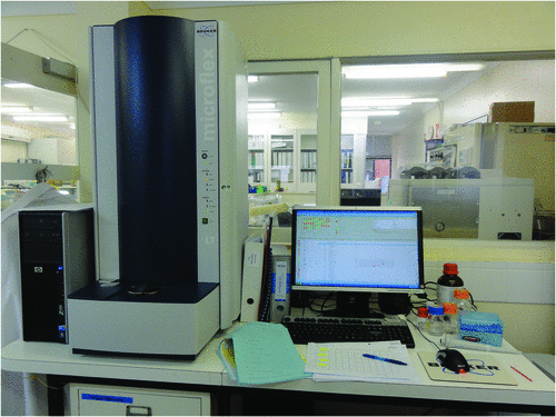
Advances in phenotypic identification have seen the advent of matrix-assisted laser desorption time of flight (MALDI-TOF) mass spectrometry (Figure 1) and to some extent this technology overcomes the constraints of the traditional methods of identifying bacteria based on the biochemical pathways of the cell. Mass spectrometry still requires the organism to be cultured, however, the identification can be performed on individual colonies growing on the primary plate. A bacterial colony is smeared within a well on a target plate and the cellular proteins are released using formic acid and a matrix solution of saturated alpha-cyano-4-hydroxycinnamic acid containing acetonitrile and trifluoroacetic acid. The high abundance ribosomal proteins between 2–20 kiloDaltons are analysed within 1–2 minutes. Identification is achieved by pattern matching of the generated protein peaks against a stored library containing spectra for 6000 species. The intensity is correlated and used for ranking results. Thus, an assigned score of >2.0 identifies the bacterium to species level, whereas a score of 1.7–2.0 identifies to genus level only (Bruker MALDI biotyper instrument, Bruker Daltonics). Although mass spectrometry has been available for protein analysis for over 50 years, it is the bioinformatics that has enabled the technology to be applied to the identification of bacteria, yeast and fungi. Only true fungi are present in the database. Oomycete fungi such as Aphanomyces and Saprolegnia that are pathogens or saprophytes in aquatic animals are not in the commercial databases at present. MALDI-TOF technology has been used for the separation of species and subspecies, strain typing, and the detection of antibiotic resistant genotypes11,12.

|
The MALDI-TOF database contains 51 of the 121 described Vibrio species, 21 of the 45 described Aeromonas species, and many other genera and species in the family Enterobacteriaceae including the exotic fish pathogen Edwardsiella ictaluri. The database also contains aquatic species from the genera Flavobacterium and Tenacibaculum (T. discolor and T. ovolyticum) but not the pathogenic species T. maritiumum, Flavobacterium columnare or F. psychrophilum. Carnobacterium maltaromaticum, a cause of pseudokidney disease in salmonids. Vagococcus fluvialis and V. lutrae are present but not the pathogens V. salmoninarum or Renibacterium salmoninarum. The database of 69 Streptococcus species does not include the human and fish pathogen Streptococcus iniae. Although MALDI-TOF identification of some bacteria is very robust, it can be unreliable for certain genera such as Vibrio and Aeromonas, and like all aspects of microbiology, the microbiologist must be aware of the limitations. The recommendation is for all unknown isolates to be tested in duplicate on the target plate. For many of the Vibrio and Aeromonas species, the same score can be obtained for duplicate spots (unpublished data) and biochemical testing or specific PCR must be done. The database tends to be more robust for human pathogens or where there are multiple strains in the database13; however, this will improve as more strains are added.
Molecular identification of many aquatic pathogens can be problematic with published PCRs for specific pathogens often cross reacting with closely related species (unpublished data). In the case of Vibrio and Aeromonas species, clonal groups or clades within these genera are so similar that at least seven housekeeping genes must be sequenced and concatenated to determine phylogenetic differences14,15. The advent of next generation sequencing technology is likely to overcome some identification problems, but at present costs are high, data storage is problematic, and the volume of data generated requires significant time to process and analyse; however, like all technology, these drawbacks will be minimised as the technology advances.
References
[1] Buller, N.B. (2014) Bacteria and Fungi from Fish and other Aquatic Animals; a practical identification manual. CABI, Oxfordshire, UK.[2] Wakabayashi, H. et al. (1986) Flexibacter maritimus sp. nov., a pathogen of marine fishes. Int. J. Syst. Bacteriol. 36, 396–398.
| Flexibacter maritimus sp. nov., a pathogen of marine fishes.Crossref | GoogleScholarGoogle Scholar |
[3] Anacker, R.L. and Ordal, E.J. (1959) Studies on the myxobacterium Chondrococcus columnaris. I. Serological typing. J. Bacteriol. 78, 25–32.
| 1:STN:280:DyaG1M7gt1KqtQ%3D%3D&md5=5f16efc9ed16389fcbcc09888e4e372bCAS | 13672906PubMed |
[4] Cipriano, R.C. and Holt, R.A. (2005) Flavobacterium psychrophilum, cause of bacterial cold-water disease and rainbow trout fry syndrome. Fish Disease leaflet No. 86. National Fish Health Research Laboratory.
[5] Fryer, J.L. and Sanders, J.E. (1981) Bacterial Kidney Disease of salmonid fish. Annu. Rev. Microbiol. 35, 273–298.
| Bacterial Kidney Disease of salmonid fish.Crossref | GoogleScholarGoogle Scholar | 1:STN:280:DyaL38%2FksVGlsA%3D%3D&md5=de940e6ad2508d1f0e6b0f4a7346947aCAS | 6794423PubMed |
[6] Cowan, S.T. and Steel, K.J. (1970) Manual for the identification of medical bacteria. Cambridge: Cambridge University Press.
[7] West, P.A. and Colwell, R.R. (1984) Identification and classification of Vibrionaceae – an overview. In Vibrios in the Environment. New York: John Wiley & Sons, pp. 285–363.
[8] Clarridge, J.E. and Zighelboim-Daum, S. (1985) Isolation and characterization of two hemolytic phenotypes of Vibrio damsela associated with a fatal wound infection. J. Clin. Microbiol. 21, 302–306.
| 1:STN:280:DyaL2M7lvVWhtw%3D%3D&md5=cf84e286b8809c99f4f19e9da0c939deCAS | 3980686PubMed |
[9] Croci, L. et al. (2007) Comparison of different biochemical and molecular methods for the identification of Vibrio parahaemolyticus. J. Appl. Microbiol. 102, 229–237.
| Comparison of different biochemical and molecular methods for the identification of Vibrio parahaemolyticus.Crossref | GoogleScholarGoogle Scholar | 1:CAS:528:DC%2BD2sXitlOmt7c%3D&md5=1026fb6e73b7828a94377132c4bd0f87CAS | 17184339PubMed |
[10] Haenen, O.L.M. et al. (2013) Bacterial infections from aquatic species: potential for and prevention of contact zoonoses. Rev. Sci. Tech. 32, 497–507.
| Bacterial infections from aquatic species: potential for and prevention of contact zoonoses.Crossref | GoogleScholarGoogle Scholar | 1:STN:280:DC%2BC2cvmtFOjtg%3D%3D&md5=823ffb474e5a6ce52a5ff9945943cb33CAS |
[11] Griffin, P.M. et al. (2012) Use of matrix-assisted laser desorption ionization – time of flight mass spectrometry to identify vancomycin-resistance Enterococci and investigate the epidemiology of an outbreak. J. Clin. Microbiol. 50, 2918–2931.
| Use of matrix-assisted laser desorption ionization – time of flight mass spectrometry to identify vancomycin-resistance Enterococci and investigate the epidemiology of an outbreak.Crossref | GoogleScholarGoogle Scholar | 1:CAS:528:DC%2BC38XhsVarsb3E&md5=4ec6026fdffd2c45a983f749abe21747CAS | 22740710PubMed |
[12] Randall, L.P. et al. (2015) Evaluation of MALDI-TOF as a method for the identification of bacteria in the veterinary diagnostic laboratory. Res. Vet. Sci. 101, 42–49.
| Evaluation of MALDI-TOF as a method for the identification of bacteria in the veterinary diagnostic laboratory.Crossref | GoogleScholarGoogle Scholar | 1:CAS:528:DC%2BC2MXhtVegur3P&md5=5da23954cc1c0c4866ff2e9835afd857CAS | 26267088PubMed |
[13] Dieckmann, R. et al. (2010) Rapid identification and characterization of Vibrio species using whole-cell MALDI-TOF mass spectrometry. J. Appl. Microbiol. 109, 199–211.
| 1:CAS:528:DC%2BC3cXpslOrsr0%3D&md5=55153df484a4a6fe3eab7d84c2ac58a1CAS | 20059616PubMed |
[14] Sawabe, T. et al. (2007) Inferring the evolutionary history of vibrios by means of multilocus sequence analysis. J. Bacteriol. 189, 7932–7936.
| Inferring the evolutionary history of vibrios by means of multilocus sequence analysis.Crossref | GoogleScholarGoogle Scholar | 1:CAS:528:DC%2BD2sXht1Knu7zK&md5=ee6c5e2d8d6fce9e5d97c7c64188f665CAS | 17704223PubMed |
[15] Martínez-Murcia, A.J. et al. (2011) Multilocus phylogenetic analysis of the genus Aeromonas. Syst. Appl. Microbiol. 34, 189–199.
| Multilocus phylogenetic analysis of the genus Aeromonas.Crossref | GoogleScholarGoogle Scholar | 21353754PubMed |
Biographies
The biography for Dr Nicky Buller is on page 103.
Sam Hair is a Microbiologist and Research Officer in the Bacteriology and Molecular Biology laboratories at Animal Health Laboratories, DAFWA. Sam is currently undertaking a PhD investigating the role of bacteria in the nutrition of post-larval abalone. Sam and Nicky work closely with pathologists and scientists at the Fish Health Unit, Department of Fisheries WA, which is co-located at DAFWA.


