Clinical and epidemiological features of congenital cytomegalovirus infection globally
Wendy J van Zuylen Serology and Virology Division
SEALS Microbiology
Prince of Wales Hospital
Randwick, NSW 2301, Australia
School of Medical Sciences
Faculty of Medicine
The University of New South Wales
Sydney, NSW 2033, Australia
Tel: +61 2 9382 9096
Fax: +61 2 9382 8533
Email: w.vanzuijlen@unsw.edu.au
Microbiology Australia 36(4) 153-156 https://doi.org/10.1071/MA15056
Published: 21 October 2015
Human cytomegalovirus (CMV) is the most common non-genetic cause of congenital disability. As a herpesvirus that infects the majority of the population, CMV is able to establish a lifelong latent infection in the host. Any time during pregnancy, a primary CMV infection, reactivation of latent CMV or a new viral strain can infect the placenta and the developing foetus, resulting in congenital CMV infection. Each year, an estimated 2000 children are born with congenital CMV infection in Australia, leaving ~500 children with permanent disabilities such as hearing or vision loss, or mental disability. Despite the clinical importance of congenital CMV, there is limited awareness and knowledge in the medical and general community about congenital CMV infection. This article reviews the global epidemiology and clinical features of maternal and congenital CMV infections.
Human CMV infection
Human CMV is a member of the Herpesviridae family of viruses, which includes Herpes simplex virus type 1 and type 2, Varicella zoster virus, Epstein-Barr virus, Human herpesvirus 6A, Human herpesvirus 6B, Human herpesvirus 7, and Human herpesvirus 81,2. The genome of human CMV is ~235 kbp and is one of the largest among the Herpesviridae3.
Human CMV infects most individuals in the world and can be acquired anytime during life: as a foetus, neonate, toddler, child or an adult. Initial infection (also known as primary infection) occurs following close personal contact. CMV is typically transmitted via body fluids, particularly breast milk, urine, genital secretions, and blood4. In addition, CMV can infect the placenta and the developing foetus5. Once infected, the human body does not clear the virus. CMV is able to persist in a latent form in either low or undetectable levels in peripheral blood mononuclear cells (CD14+) and bone marrow cells (CD34+ and CD33+)6. Stimuli such as inflammation, immune impairment due to pregnancy, medical treatment with immunomodulating agents such as corticosteroids, chemotherapy, and immunosuppressive therapy post organ transplantation may stimulate reactivation and growth of latent CMV7. Considering CMV secretion in urine and cervical-vaginal fluids increases during pregnancy with increasing gestational age, hormonal changes related to pregnancy may also stimulate reactivation of CMV8.
Epidemiology of maternal and congenital CMV infections
CMV is a common cause of infections worldwide. Antibodies to CMV, representing a previous infection, can be detected in 45 to >90% of women of reproductive age9. The percentage of women that are infected with CMV varies between countries and tends to be the lowest in Western Europe, Australia, Canada and the United States and the highest in South America, Africa and Asia9. Particularly, in Australia, the average seroprevalence rate of CMV for women between the ages of 14 to 44 years is 58%10. However, even within countries the rate of CMV infected women varies by socio-economic status and ethnicity9,11.
Approximately 1–2% of initially uninfected pregnant women will acquire CMV by the time of delivery12. A possible source of CMV for these women is young children whose saliva and urine contain high levels of CMV13. In addition, a partner who is infected with CMV is an additional possible risk factor for infection during pregnancy, as CMV is present in semen, and can be transmitted sexually12. Among the women who acquire a primary infection during pregnancy 32% transmit CMV to the foetus via the placenta, resulting in congenital CMV (cCMV) infection14. Only a percentage of cCMV infected children will exhibit symptoms at birth or develop CMV associated symptoms later in life, as further described in detail below.
The foetus can also be infected by a woman’s latent virus or re-infection with a different strain of CMV (secondary infection)15. The risk of transmitting CMV to the foetus is reported to be higher when a pregnant woman acquires a primary infection during the first half of the pregnancy compared to secondary infections, or infection in the second half of pregnancy16. Kenneson14 reported 1.4% of secondary infections lead to foetal infection. However, considering the high seroprevalence of CMV, it is estimated that more than two-thirds of CMV infected children are born to mothers who were already infected with CMV17.
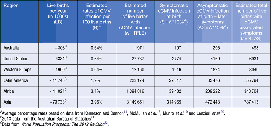
Intrauterine CMV infection occurs in 0.2 to 2% (average of 0.64%) of live births in the Unites States, Australia and Western Europe (Table 1)14,18–20. In addition, the limited studies of regions in Latin America, Africa, and Asia have reported a birth prevalence of cCMV infection ranging from 0.6 to 6.1% of pregnancies20. Based on the number of live births per year22 and reported cCMV prevalence14,18–20, this translates to an estimated ~0.12 million cCMV infections in developed countries per year, and ~0.7 million to 4.5 million cCMV infections annually in developing countries. Particularly in Australia, an estimated ~2000 children are born with cCMV infection in Australia each year (Table 1). Nonetheless, in practice, most congenital CMV infections remain undiagnosed18.

|
Clinical features of maternal CMV infection
The majority of CMV infections in immunocompetent individuals do not cause symptoms; however, clinical manifestations could include glandular fever (mononucleosis) syndrome characterised by flu-like symptoms, or occasionally persistent fever24. Several studies reported that pregnant women, who acquired a primary CMV infection, experienced mononucleosis, fever, fatigue, and headache25. Additionally, Nigro24 observed a significantly higher number of pregnant women with primary CMV infection presenting with symptoms compared to pregnant women with recurrent or latent CMV infection. A review of congenital CMV cases in Australia reported more than half of the mothers had evidence of, or could recall experiencing symptoms of fever during pregnancy26. In addition to clinical symptoms, laboratory examination may show an increase in lymphocytes in the blood and increased serum levels of liver enzymes (alanine transaminase and aspartate transaminase)24. Since all of these clinical manifestations are not only observed upon a CMV infection, they do not represent specific indicators of maternal CMV infection. However, collection of the clinical history and laboratory examination may be extremely useful for dating the onset of infection to determine the risk of CMV transmission to the foetus and risk of cCMV disease.
Clinical features of congenital cytomegalovirus disease
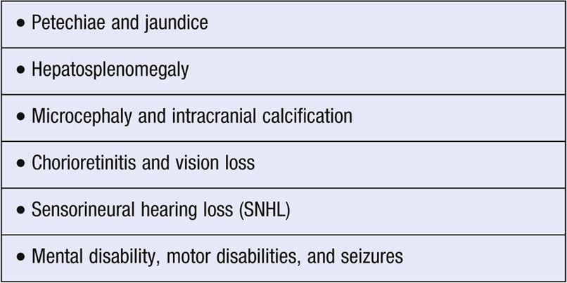
A minority (~10%) of cCMV infected children present symptoms at birth (Table 2). Physical signs such as petechiae, jaundice, and hepatosplenomegaly are common and have been observed in 28 to 50% of children with cCMV infection18,27. Neurological abnormality, including microcephaly and intracranial calcification has been reported to occur in 18–38% of cCMV infected children. The majority of these affected children develop sensorineural hearing loss, mental disability, motor deficits, chorioretinitis and seizures16,23,26.

|
A significant amount (~15%) of initially asymptomatic CMV infected children will encounter developmental difficulties, neurological problems, or hearing loss before the age of five9,18,28. Among those with hearing loss ~40% of children may develop severe to profound impairment of both ears23. Other neurological complications such as microcephaly, neuromuscular defects, and chorioretinitis may also develop in initially asymptomatic CMV infected children, but at a lower rate compared to symptomatic infection1.
Congenital CMV infection may also result in adverse pregnancy outcomes, as cCMV has been associated with fetal death in utero, neonatal death, preterm birth and maternal pregnancy complications, including preeclampsia29–33.
Concluding remarks
CMV continues to be the leading infectious cause of congenital malformation in developed countries. More children may be affected by cCMV than by any other childhood disorder, such as down syndrome, fetal alcohol syndrome, and spina bifida. Each year in Australia, an estimated 2000 children are born with cCMV infection, leaving ~500 children with permanent disabilities such as hearing or vision loss, or mental disability. Even though the rates of maternal and cCMV infection are still lacking for many parts of the world, which likely underestimates the global impact of cCMV infection, the importance of cCMV infection and disease as a large public health problem is self-evident.
References
[1] Manicklal, S. et al. (2013) The ‘silent’ global burden of congenital cytomegalovirus. Clin. Microbiol. Rev. 26, 86–102.| The ‘silent’ global burden of congenital cytomegalovirus.Crossref | GoogleScholarGoogle Scholar | 1:CAS:528:DC%2BC3sXntVyms7w%3D&md5=67d112e96f469983b3480909f453423cCAS | 23297260PubMed |
[2] Crough, T. and Khanna, R. (2009) Immunobiology of human cytomegalovirus: from bench to bedside. Clin. Microbiol. Rev. 22, 76–98.
| Immunobiology of human cytomegalovirus: from bench to bedside.Crossref | GoogleScholarGoogle Scholar | 1:CAS:528:DC%2BD1MXnslKrs7o%3D&md5=3e95a9b19b778cbc193b76de479ed5a3CAS | 19136435PubMed |
[3] Davison, A.J. (2013) Comparitive genomics of primate cytomegaloviruses. In Cytomegaloviruses: From Molecular Pathogenesis to Intervention (Reddehase, M.J., ed.), pp. 1–22, Caister Academic Press.
[4] Cannon, M.J. et al. (2011) Review of cytomegalovirus shedding in bodily fluids and relevance to congenital cytomegalovirus infection. Rev. Med. Virol. 21, 240–255.
| Review of cytomegalovirus shedding in bodily fluids and relevance to congenital cytomegalovirus infection.Crossref | GoogleScholarGoogle Scholar | 1:CAS:528:DC%2BC3MXotVentr4%3D&md5=b7dafbcb5f37f99b05fe30fec9e88cb6CAS | 21674676PubMed |
[5] Fisher, S. et al. (2000) Human cytomegalovirus infection of placental cytotrophoblasts in vitro and in utero: implications for transmission and pathogenesis. J. Virol. 74, 6808–6820.
| Human cytomegalovirus infection of placental cytotrophoblasts in vitro and in utero: implications for transmission and pathogenesis.Crossref | GoogleScholarGoogle Scholar | 1:CAS:528:DC%2BD3cXkvFCmt78%3D&md5=149f24ac07c6790e8fa3cddeb726b792CAS | 10888620PubMed |
[6] Slobedman, B. et al. (2010) Human cytomegalovirus latent infection and associated viral gene expression. Future Microbiol. 5, 883–900.
| Human cytomegalovirus latent infection and associated viral gene expression.Crossref | GoogleScholarGoogle Scholar | 1:CAS:528:DC%2BC3cXmvFeju7Y%3D&md5=d8147aac39f1aa1ec6d52db7ef5957b0CAS | 20521934PubMed |
[7] Mocarski, E. et al. (2007) Cytomegaloviruses. In Fields Virology (5th edn) (Knipe, D., et al., eds), Lippincott Williams & Wilkins.
[8] Knowles, W.A. et al. (1982) A comparison of cervical cytomegalovirus (CMV) excretion in gynaecological patients and post-partum women. Arch. Virol. 73, 25–31.
| A comparison of cervical cytomegalovirus (CMV) excretion in gynaecological patients and post-partum women.Crossref | GoogleScholarGoogle Scholar | 1:STN:280:DyaL3s%2FitFOrtg%3D%3D&md5=2f580e19a19224cbfddad7d509708333CAS | 6289775PubMed |
[9] Cannon, M.J. et al. (2010) Review of cytomegalovirus seroprevalence and demographic characteristics associated with infection. Rev. Med. Virol. 20, 202–213.
| Review of cytomegalovirus seroprevalence and demographic characteristics associated with infection.Crossref | GoogleScholarGoogle Scholar | 20564615PubMed |
[10] Seale, H. et al. (2006) National serosurvey of cytomegalovirus in Australia. Clin. Vaccine Immunol. 13, 1181–1184.
| National serosurvey of cytomegalovirus in Australia.Crossref | GoogleScholarGoogle Scholar | 1:CAS:528:DC%2BD28Xht1Gksb7N&md5=fbc1f65aaf155c066d05408862d06197CAS | 16957061PubMed |
[11] Basha, J. et al. (2014) Congenital cytomegalovirus infection is associated with high maternal socio-economic status and corresponding low maternal cytomegalovirus seropositivity. J. Paediatr. Child Health 50, 368–372.
| Congenital cytomegalovirus infection is associated with high maternal socio-economic status and corresponding low maternal cytomegalovirus seropositivity.Crossref | GoogleScholarGoogle Scholar | 24593837PubMed |
[12] Hyde, T.B. et al. (2010) Cytomegalovirus seroconversion rates and risk factors: implications for congenital CMV. Rev. Med. Virol. 20, 311–326.
| Cytomegalovirus seroconversion rates and risk factors: implications for congenital CMV.Crossref | GoogleScholarGoogle Scholar | 20645278PubMed |
[13] Adler, S.P. (1991) Cytomegalovirus and child day care: risk factors for maternal infection. Pediatr. Infect. Dis. J. 10, 590–594.
| Cytomegalovirus and child day care: risk factors for maternal infection.Crossref | GoogleScholarGoogle Scholar | 1:STN:280:DyaK3MzmslGktA%3D%3D&md5=f51d109471bc437fc76d342238a44742CAS | 1653939PubMed |
[14] Kenneson, A. and Cannon, M.J. (2007) Review and meta-analysis of the epidemiology of congenital cytomegalovirus (CMV) infection. Rev. Med. Virol. 17, 253–276.
| Review and meta-analysis of the epidemiology of congenital cytomegalovirus (CMV) infection.Crossref | GoogleScholarGoogle Scholar | 17579921PubMed |
[15] Wang, C. et al. (2011) Attribution of congenital cytomegalovirus infection to primary versus non-primary maternal infection. Clin. Infect. Dis. 52, e11–e13.
| Attribution of congenital cytomegalovirus infection to primary versus non-primary maternal infection.Crossref | GoogleScholarGoogle Scholar | 21288834PubMed |
[16] Pass, R.F. et al. (2006) Congenital cytomegalovirus infection following first trimester maternal infection: symptoms at birth and outcome. J. Clin. Virol. 35, 216–220.
| Congenital cytomegalovirus infection following first trimester maternal infection: symptoms at birth and outcome.Crossref | GoogleScholarGoogle Scholar | 16368262PubMed |
[17] de Vries, J.J. et al. (2013) The apparent paradox of maternal seropositivity as a risk factor for congenital cytomegalovirus infection: a population-based prediction model. Rev. Med. Virol. 23, 241–249.
| The apparent paradox of maternal seropositivity as a risk factor for congenital cytomegalovirus infection: a population-based prediction model.Crossref | GoogleScholarGoogle Scholar | 23559569PubMed |
[18] McMullan, B.J. et al. (2011) Congenital cytomegalovirus – time to diagnosis, management and clinical sequelae in Australia: opportunities for earlier identification. Med. J. Aust. 194, 625–629.
| 21692718PubMed |
[19] Munro, S.C. et al. (2005) Diagnosis of and screening for cytomegalovirus infection in pregnant women. J. Clin. Microbiol. 43, 4713–4718.
| Diagnosis of and screening for cytomegalovirus infection in pregnant women.Crossref | GoogleScholarGoogle Scholar | 1:CAS:528:DC%2BD2MXhtVOrtLbK&md5=251d466089be2daf14860b6bf7fb9809CAS | 16145132PubMed |
[20] Lanzieri, T.M. et al. (2014) Systematic review of the birth prevalence of congenital cytomegalovirus infection in developing countries. Int. J. Infect. Dis. 22, 44–48.
| Systematic review of the birth prevalence of congenital cytomegalovirus infection in developing countries.Crossref | GoogleScholarGoogle Scholar | 24631522PubMed |
[21] Australian Bureau of Statistics (2013) Births, Australia 2013, http://www.abs.gov.au/AUSSTATS/abs@.nsf/mf/3301.0 (accessed July 2015).
[22] UN. (2012) World Population Prospects, The 2012 Revision, http://esa.un.org/wpp/Excel-Data/fertility.htm (accessed July 2015).
[23] Goderis, J. et al. (2014) Hearing loss and congenital CMV infection: a systematic review. Pediatrics 134, 972–982.
| Hearing loss and congenital CMV infection: a systematic review.Crossref | GoogleScholarGoogle Scholar | 25349318PubMed |
[24] Nigro, G. et al. (2003) Clinical manifestations and abnormal laboratory findings in pregnant women with primary cytomegalovirus infection. BJOG 110, 572–577.
| Clinical manifestations and abnormal laboratory findings in pregnant women with primary cytomegalovirus infection.Crossref | GoogleScholarGoogle Scholar | 12798474PubMed |
[25] Revello, M.G. and Gerna, G. (2002) Diagnosis and management of human cytomegalovirus infection in the mother, fetus, and newborn infant. Clin. Microbiol. Rev. 15, 680–715.
| Diagnosis and management of human cytomegalovirus infection in the mother, fetus, and newborn infant.Crossref | GoogleScholarGoogle Scholar | 12364375PubMed |
[26] Munro, S.C. et al. (2005) Symptomatic infant characteristics of congenital cytomegalovirus disease in Australia. J. Paediatr. Child Health 41, 449–452.
| Symptomatic infant characteristics of congenital cytomegalovirus disease in Australia.Crossref | GoogleScholarGoogle Scholar | 16101982PubMed |
[27] Kylat, R.I. et al. (2006) Clinical findings and adverse outcome in neonates with symptomatic congenital cytomegalovirus (SCCMV) infection. Eur. J. Pediatr. 165, 773–778.
| Clinical findings and adverse outcome in neonates with symptomatic congenital cytomegalovirus (SCCMV) infection.Crossref | GoogleScholarGoogle Scholar | 16835757PubMed |
[28] Dahl, H.H. et al. (2013) Etiology and audiological outcomes at 3 years for 364 children in Australia. PLoS One 8, e59624.
| Etiology and audiological outcomes at 3 years for 364 children in Australia.Crossref | GoogleScholarGoogle Scholar | 1:CAS:528:DC%2BC3sXmtVensbo%3D&md5=251c142629fc5500c9bff93de5a469d8CAS | 23555729PubMed |
[29] Iwasenko, J.M. et al. (2011) Human cytomegalovirus infection is detected frequently in stillbirths and is associated with fetal thrombotic vasculopathy. J. Infect. Dis. 203, 1526–1533.
| Human cytomegalovirus infection is detected frequently in stillbirths and is associated with fetal thrombotic vasculopathy.Crossref | GoogleScholarGoogle Scholar | 21592980PubMed |
[30] Pereira, L. et al. (2014) Intrauterine growth restriction caused by underlying congenital cytomegalovirus infection. J. Infect. Dis. 209, 1573–1584.
| Intrauterine growth restriction caused by underlying congenital cytomegalovirus infection.Crossref | GoogleScholarGoogle Scholar | 1:CAS:528:DC%2BC2cXnt1CmtLg%3D&md5=fe270b2b7d97aa348ac72318112cd191CAS | 24403553PubMed |
[31] Williams, E.J. et al. (2013) Viral infections: contributions to late fetal death, stillbirth, and infant death. J. Pediatr. 163, 424–428.
| Viral infections: contributions to late fetal death, stillbirth, and infant death.Crossref | GoogleScholarGoogle Scholar | 23507026PubMed |
[32] Lorenzoni, F. et al. (2014) Neonatal screening for congenital cytomegalovirus infection in preterm and small for gestational age infants. J. Matern. Fetal Neonatal Med. 27, 1589–1593.
| 1:STN:280:DC%2BC2c3lvVOjtg%3D%3D&md5=c7a7b008b84953d231c414a1133f1c7bCAS | 24328547PubMed |
[33] Xie, F. et al. (2010) An association between cytomegalovirus infection and pre-eclampsia: a case-control study and data synthesis. Acta Obstet. Gynecol. Scand. 89, 1162–1167.
| An association between cytomegalovirus infection and pre-eclampsia: a case-control study and data synthesis.Crossref | GoogleScholarGoogle Scholar | 20804342PubMed |
Biography
Wendy van Zuylen is a postdoctoral scientist in the School of Medical Sciences, Faculty of Medicine at the University of New South Wales. Her current research at the Virology Research Laboratory aims to understand the pathogenesis of congenital cytomegalovirus infection.


