First report of Septoria leaf spot of pistachio in Iran
M. A. Aghajani A D , B. Aghapour B and T. J. Michailides CA Department of Plant Protection Research, Agricultural and Natural Resources Research Center of Golestan Province, Gorgan, Iran.
B Department of Plant Protection, Faculty of Agriculture and Natural Resources, University of Tehran, Karaj, Iran.
C Department of Plant Pathology, University of California, Davis/ Kearney Agricultural Center, Parlier 93648, USA.
D Corresponding author. Email: maaghajanina@yahoo.com
Australasian Plant Disease Notes 4(1) 29-31 https://doi.org/10.1071/DN09012
Submitted: 5 November 2008 Accepted: 12 March 2009 Published: 20 April 2009
Abstract
During the summer of 2008, a severe foliar disease was observed on pistachio trees in Golestan Province, in northern Iran. Signs and symptoms of the disease were abundant pycnidia followed by brown spots on leaves. Septoria pistacina was consistently isolated from all the diseased leaves. This is the first report of Septoria leaf spot on pistachio in Iran.
Pistachio (Pistacia vera) has a recorded history of more than 800 years in Golestan Province, northern Iran. Wild populations of this species are distributed over a 40 000 ha area in the northern and eastern regions of this Province. Despite this, only ~30 ha of improved varieties have been established in this region (Karimidoost 2001).
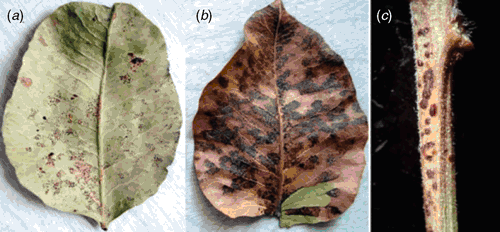
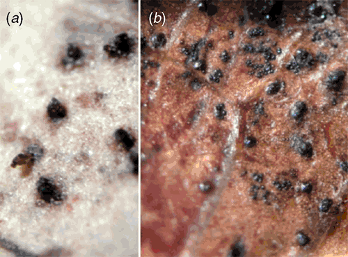
During the early summer (July) of 2008, symptoms of a leaf spot disease were observed in several pistachio orchards. At first dark pycnidia appeared on both sides of the leaf. The pycnidia were scattered over a circular area ~0.5 cm or less in diameter (Fig. 1a). Later, more pycnidia were formed and this area enlarged to more than 1 cm in diameter. As the pycnidia matured, cirrhi of pycnidiospores could be seen extruding from the ostioles, which made the leaf tissue areas bearing pycnidia very prominent (Fig. 2a). These areas with pycnidia often coalesced to form larger lesions, sometimes occupying almost the entire leaf. Spermagonia of the fungus were produced on both sides of the leaf, among the pycnidia (Fig. 2b). They were immersed, tiny and dark. Only late in the season, the leaf tissue bearing pycnidia and spermagonia became chlorotic and eventually necrotic (Fig. 1b). Similar symptoms were observed on the petioles (Fig. 1c).

|

|
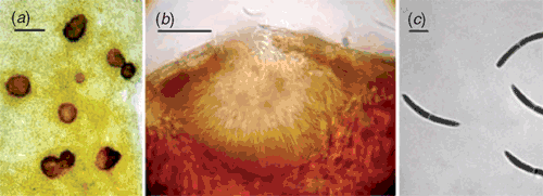
The pycnidia, immersed in the leaf tissue, were tan-brown, globose-depressed to lens-shaped and ranged from 80–175 µm (height) to 125–325 µm (width) (Fig. 3b). The pycnidiospores were hyaline, filiform, curved, and almost falcate, with obtuse ends, and usually one septum at their middle where they were constricted (Fig. 3c). Pycnidiospores with 2 or 3 septa were rarely observed (Table 1) and measured 25–47 × 2.5–5 µm.

|

|
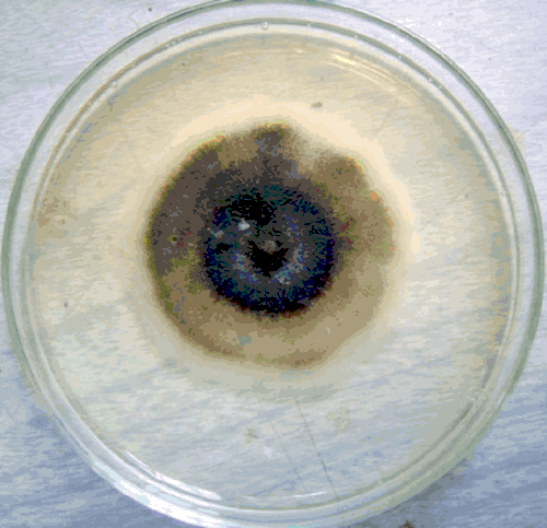
To determine the causal agent of the disease, samples of diseased plants were collected from several locations. Infected tissues were placed in moist chambers until cirrhi of pycnidiospores extruded from the ostioles of pycnidia. Small pieces of cirrhi were placed on 2% water agar, and after 2 days, germinated single pycnidiospores were transferred to potato dextrose agar and incubated at 25°C in darkness. Mycelial growth of the fungus under these conditions was very slow. The fungal colony was initially white, but after 7–10 days incubation became dark grey (Fig. 4). After this time, globose to sub-globose spermagonia (52.5–107.5 (height) × 40–92.5 µm (width)) started forming on the culture plate (Fig. 3a). No pycnidia were formed in these cultures after incubation of the Petri plates for a month.

|
The causal fungus was identified as Septoria pistacina Allesch., based on morphological characteristics of the fungus and type of signs and symptoms on pistachio leaves (Chitzanidis 1956). It is easily separated from the two other Septoria species reported on pistachio in the laboratory, based on morphological characteristics and number of septa of pycnidiospores (Table 1).
Three species of the genus Septoria have been reported on pistachio worldwide, S. pistaciarum, S. pistaciae and S. pistacina (Teviotdale et al. 2001). Septoria leaf spot (caused by S. pistaciarum) was detected in the United States for the first time in 1964 within an experimental pistachio planting at Brownwood, Texas (Young and Michailides 1989; Call and Matheron 2000). The first observation of the disease in Arizona pistachio trees did not occur until 1986 (Young and Michailides 1989). Eskalen et al. (2001) reported it as the causul agent of Septoria leaf spot from pistachio trees in East-Mediterranean and South-east Anatolian regions. Septoria pistaciae was reported from the United States (California), Asia (Armenia, Republic of Georgia, India, Israel, Kazakhstan, Kirgizstan, Syria, Turkey, Turkmenistan and Uzbekistan), Europe (Albania, France, Greece, Italy, Portugal and Ukraine), and Africa (Egypt) (Pantidou 1973; Dudka et al. 2004; Haggag et al. 2006).
Septoria pistacina is reported from Greece (Chitzanidis 1956; Spaulding 1961), Syria (Spaulding 1961) and Turkey (T. J. Michailides, unpubl. data). It appears to have a limited distribution, compared with the other two species of Septoria on pistachio. Septoria pistacina is apparently limited to the Mediterranean countries, and our report for its occurrence in Iran, further documents its origin in this region. To our knowledge, this is the first report of Septoria leaf spot of pistachio caused by S. pistacina in Iran.
Chitzanidis A
(1956) Species of Septoria on the leaves of Pistacia vera L., and their perfect stages. Annales of Institut Phytopathologique Benaki 10, 29–44.

Haggag WM,
Abou Rayya MSM, Kasim NE
(2006) First Report of Septoria pistaciae Causing Leaf Spot of Pistachio in Egypt. Plant Disease 90, 1553.
| Crossref | GoogleScholarGoogle Scholar |

Young DJ, Michailides TJ
(1989) First report of Septoria leaf spot of pistachio in Arizona. Plant Disease 73, 775.
| Crossref | GoogleScholarGoogle Scholar |


