Activated sludge foaming: can phage therapy provide a control strategy?
Steve Petrovski A B and Robert Seviour A CA Department of Physiology, Anatomy and Microbiology, La Trobe University, Bundoora, Vic. 3086, Australia
B Email: steve.petrovski@latrobe.edu.au
C Email: r.seviour@latrobe.edu.au
Microbiology Australia 39(3) 162-164 https://doi.org/10.1071/MA18048
Published: 7 August 2018
Foaming in activated sludge systems is a global problem leading to environmental, cosmetic and operational problems. Proliferation of filamentous hydrophobic bacteria (including the Mycolata) are responsible for the stabilisation of foams. Currently no reliable methods exist to control these. Reducing the levels of the filamentous bacteria with bacteriophages below the threshold supporting foaming is an attractive approach to control their impact. We have isolated 88 bacteriophages that target members of the foaming Mycolata. These double stranded DNA phages have been characterised and are currently being assessed for their performance as antifoam agents.
The activated sludge process
The activated sludge process is a robust and proven system for treating domestic and industrial wastewater and is used globally1. It relies on a specialised community of microbes organised into structures called flocs, which metabolise organic nutrients and remove inorganic nitrogen and phosphorus compounds so that the treated effluent can be discharged safely into a receiving body of water without leading to eutrophication from growth of toxic Cyanobacteria1.
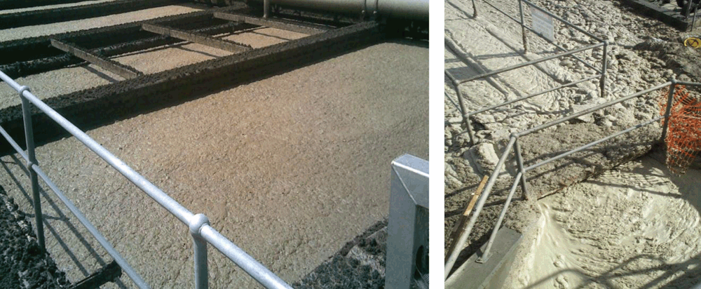
These systems are no longer considered as wastewater water disposal systems, but as valuable sources of purified water for reuse and useful chemicals. Despite their popularity most suffer from the problem of foaming where a brown foam layer develops on the reactor surface and leaves in the treated effluent2.
What is foaming?
Foaming, which increases plant operating costs, reduces effluent quality and acts as a source of opportunistic human pathogens, is a flotation event, requiring three components; air bubbles, surfactants and hydrophobic particles (bacterial cells), which act to stabilise it (Figure 1). With only air bubbles and surfactants, an unstable foam forms, and is often seen in start-up, where abundances of hydrophobic bacteria are below the threshold supporting foam formation3. With insufficient levels of surfactants, air bubbles collapse and a greasy surface layer, a scum, forms, consisting of hydrophobic bacteria. There are no reliable control measures to deal with an already established foam, but any proposed strategy should target the hydrophobic bacterial cells, since control of the other two is impractical.
It is now clear that a diverse range of bacteria are responsible for foaming episodes4–8. Theoretically, any sufficiently hydrophobic cell can stabilise this foam, but surveys suggest that the unbranched actinobacterial filamentous organism ‘Microthrix parvicella’ and the right angled branching mycolic acid producing filaments placed in the Mycolata (include members of the genus Gordonia, Nocardia, Rhodococcus, Tsukamurella, Skermania and related members)5,7 are the main culprits (Figure 2). Being strongly hydrophobic, these organisms escape the plant bulk liquid to the air liquid interface, often carrying biomass or sludge with them, and there attach to the liquid air films of the bubbles, preventing liquid drainage from them and hence stabilise them.
Foaming control
The conventional way to deal with existing foams is commonly non-selective using bactericidal chemicals, where organisms other than those causing foaming are likely to be harmed. Others include changing aeration rates, or reducing sludge ages hoping that foaming organisms, assumed to be slow growing, are washed out. Unfortunately other desirable bacteria are also lost. All reflect our inadequate understanding of the microbial ecology of foams.
What is needed is a specific control strategy, which is environmentally safe and importantly only removes the nuisance organisms9,10. Bacteriophages (or phages), viruses that specifically target only their bacterial hosts, and are naturally occurring and self-dosing, seem especially attractive. They are used clinically to treat infectious bacterial diseases, where the causative organism is antibiotic resistant11. As they infect their hosts, they replicate and upon lysis, release often hundreds of new phages that then infect other host cells.
The general experience has been that wherever bacteria are present, phages able to lyse them will also be present. Consequently, phages lytic for members of the foaming Mycolata should be plentiful in activated sludge. Thomas and colleagues9, demonstrated that phages, some polyvalent, are isolated readily from activated sludge plants, capable of killing their Mycolata hosts under laboratory conditions. What we know of phage/host population dynamics suggest that their presence would not lead to the total loss of their bacterial hosts. Such outcomes would be disastrous, since Mycolata play important roles in metabolising recalcitrant xenobiotics there. Strategically the aim is to reduce Mycolata numbers below individual threshold levels needed for stable foam formation. This requires identifying which are the causative organisms. FISH probing provides the tools to screen foam samples, and while their true level of biodiversity, is not known, FISH data suggest a limited number of foaming bacteria are common in plants.
What have we achieved?
The advent of Next Generation DNA Sequencing (NGS) has revolutionised our understanding of phage genomics and allowed us to screen those attractive for phage therapy, avoiding those possessing virulence or toxin genes. We have isolated 88 double stranded DNA phages that seem suitable for further study. These include phages against foaming Gordonia12,13, Rhodococcus14–16, Nocardia17, Skermania18 and Tsukamurella19,20. While most are monovalent, polyvalent phages are clearly more attractive, since most foams contain more than one Mycolata member. Not surprisingly, sequencing reveals that all are highly novel at the DNA level, but share the same genomic arrangements. They have all been screened against foaming Mycolata hosts using a simple foaming apparatus21. Almost always their foaming abilities were reduced to the point where no foam was detected as previously described13,22.
Where next?
The next step is to scale up the system. Before this is warranted, it is important to determine their host specificities and burst sizes in situ, their persistence times in full scale plants, the host cell threshold values for foam production and how much inoculum is required. Equally, the location for introducing the phages into the system is likely to be important. These parameters are plant specific, and so will need to be determined on an individual basis. Whether these phages are involved in gene transfer between host cells (transduction), and whether they acquire, as a consequence, antibiotic resistant genes and hence pose a possible threat upon release into the environment, will need investigation. In addition, the possibility of the bacterial strains developing phage resistance will need to be investigated and one possible solution would be to add multiple phages for the same host as multiple mutations is less likely.
References
[1] Seviour, R. and Nielsen, P.H. (2010) Microbial ecology of activated sludge. IWA publishing.[2] Jenkins, D. et al. (2003) Manual on the causes and control of activated sludge bulking, foaming, and other solids separation problems. CRC Press.
[3] Petrovski, S. et al. (2011) An examination of the mechanism for stable foam formation in activated sludge systems. Water Res. 45, 2146–2154.
[4] Soddell, J. (1998) Foaming. In The microbiology of activated sludge. pp. 161–202, Kluwer Academic Publishers, Dordrecht.
[5] de los Reyes III (2010) Foaming. In The Microbial Ecology of Activated Sludge. pp. 215–258. IWA Publishing, London.
[6] Seviour, R.J. et al. (2008) Ecophysiology of the Actinobacteria in activated sludge systems. Antonie van Leeuwenhoek 94, 21–33.
| Ecophysiology of the Actinobacteria in activated sludge systems.Crossref | GoogleScholarGoogle Scholar |
[7] Nielsen, P.H. et al. (2009) Identity and physiology of filamentous bacteria in activated sludge. FEMS Microbiol. Rev. 33, 969–998.
[8] Kragelund, C. et al. (2007) Ecophysiology of mycolic acid-containing Actinobacteria (Mycolata) in activated sludge foams. FEMS Microbiol. Ecol. 61, 174–184.
| Ecophysiology of mycolic acid-containing Actinobacteria (Mycolata) in activated sludge foams.Crossref | GoogleScholarGoogle Scholar |
[9] Thomas, J.A. et al. (2002) Fighting foam with phages? Water Sci. Technol. 46, 511–518.
| Fighting foam with phages?Crossref | GoogleScholarGoogle Scholar |
[10] Liu, M. et al. (2015) Bacteriophages of wastewater foaming-associated filamentous Gordonia reduce host levels in raw activated sludge. Sci. Rep. 5, 13754.
| Bacteriophages of wastewater foaming-associated filamentous Gordonia reduce host levels in raw activated sludge.Crossref | GoogleScholarGoogle Scholar |
[11] Abedon, S.T. et al. (2017) Phage therapy:past, present and future. Front. Microbiol. 8, 981.
| Phage therapy:past, present and future.Crossref | GoogleScholarGoogle Scholar |
[12] Dyson, Z.A. et al. (2015) Lysis to kill: evaluation of the lytic abilities, and genomics of nine bacteriophages infective for Gordonia spp. and their potential use in activated sludge foam biocontrol. PLoS One 10, e0134512.
| Lysis to kill: evaluation of the lytic abilities, and genomics of nine bacteriophages infective for Gordonia spp. and their potential use in activated sludge foam biocontrol.Crossref | GoogleScholarGoogle Scholar |
[13] Petrovski, S. et al. (2011) Characterization of the genome of the polyvalent lytic bacteriophage GTE2, which has potential for biocontrol of Gordonia-, Rhodococcus-, and Nocardia-stabilized foams in activated sludge plants. Appl. Environ. Microbiol. 77, 3923–3929.
| Characterization of the genome of the polyvalent lytic bacteriophage GTE2, which has potential for biocontrol of Gordonia-, Rhodococcus-, and Nocardia-stabilized foams in activated sludge plants.Crossref | GoogleScholarGoogle Scholar |
[14] Petrovski, S. et al. (2012) Small but sufficient: the Rhodococcus phage RRH1 has the smallest known Siphoviridae genome at 14.2 kilobases. J. Virol. 86, 358–363.
| Small but sufficient: the Rhodococcus phage RRH1 has the smallest known Siphoviridae genome at 14.2 kilobases.Crossref | GoogleScholarGoogle Scholar |
[15] Petrovski, S. et al. (2013) Genome sequence and characterization of a Rhodococcus equi phage REQ1. Virus Genes 46, 588–590.
| Genome sequence and characterization of a Rhodococcus equi phage REQ1.Crossref | GoogleScholarGoogle Scholar |
[16] Petrovski, S. et al. (2013) Characterization and whole genome sequences of the Rhodococcus bacteriophages RGL3 and RER2. Arch. Virol. 158, 601–609.
| Characterization and whole genome sequences of the Rhodococcus bacteriophages RGL3 and RER2.Crossref | GoogleScholarGoogle Scholar |
[17] Petrovski, S. et al. (2014) Genome sequence of the Nocardia bacteriophage NBR1. Arch. Virol. 159, 167–173.
| Genome sequence of the Nocardia bacteriophage NBR1.Crossref | GoogleScholarGoogle Scholar |
[18] Dyson, Z.A. et al. (2016) Isolation and characterization of bacteriophage SPI1, which infects the activated-sludge-foaming bacterium Skermania piniformis. Arch. Virol. 161, 149–158.
| Isolation and characterization of bacteriophage SPI1, which infects the activated-sludge-foaming bacterium Skermania piniformis.Crossref | GoogleScholarGoogle Scholar |
[19] Dyson, Z.A. et al. (2015) Three of a Kind: Genetically Similar Tsukamurella Phages TIN2, TIN3, and TIN4. Appl. Environ. Microbiol. 81, 6767–6772.
| Three of a Kind: Genetically Similar Tsukamurella Phages TIN2, TIN3, and TIN4.Crossref | GoogleScholarGoogle Scholar |
[20] Petrovski, S. et al. (2011) Genome sequence and characterization of the Tsukamurella bacteriophage TPA2. Appl. Environ. Microbiol. 77, 1389–1398.
| Genome sequence and characterization of the Tsukamurella bacteriophage TPA2.Crossref | GoogleScholarGoogle Scholar |
[21] Blackall, L.L. and Marshall, K.C. (1989) The mechanism of stabilization of actinomycete foams and the prevention of foaming under laboratory conditions. J. Ind. Microbiol. 4, 181–187.
| The mechanism of stabilization of actinomycete foams and the prevention of foaming under laboratory conditions.Crossref | GoogleScholarGoogle Scholar |
[22] Petrovski, S. et al. (2011) Prevention of Gordonia and Nocardia stabilized foam formation by using bacteriophage GTE7. Appl. Environ. Microbiol. 77, 7864–7867.
| Prevention of Gordonia and Nocardia stabilized foam formation by using bacteriophage GTE7.Crossref | GoogleScholarGoogle Scholar |
Biographies
Dr Steve Petrovski is a Senior Lecturer at La Trobe University. His research interests include mobile genetic elements, bacteriophages and their applications and microbial genetics.
Professor Robert Seviour is an Emeritus Professor at La Trobe University. His research interests include wastewater microbiology, bacteriophage biocontrol and fungal polysaccharides.


