First report of powdery mildew caused by anamorphic Podosphaera sp. on Coreopsis sp. in India
Pankaj Baiswar A B , Satish Chandra A , T. K. Bag A and S. V. Ngachan AA ICAR Research Complex for NEH Region, Umiam-793103, Meghalaya, India.
B Corresponding author. Email: pbaiswar@yahoo.com
Australasian Plant Disease Notes 5(1) 90-92 https://doi.org/10.1071/DN10032
Submitted: 12 July 2010 Accepted: 16 August 2010 Published: 30 August 2010
Abstract
This is the first report of powdery mildew on leaves of Coreopsis sp., an ornamental plant, caused by the anamorph of Podosphaera sp. in India.
Coreopsis belongs to the Asteraceae and is the largest genus in the tribe Coreopsideae. It is used as an ornamental plant in India. African species of Coreopsis are now considered as belonging to the genus Bidens. Coreopsis is now regarded as a New World genus in which the evolution of morphologically useful taxonomic characters has been complex and their value compromised for delimitation of monophyletic groups (Crawford and Mort 2005).
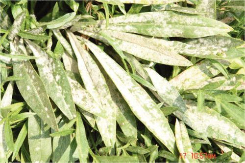
Powdery mildew symptoms were observed in April 2010 on almost 80% of the Coreopsis sp. plants grown in a garden at Umsaw Khawan (RiBhoi, Meghalaya, India). Symptoms included circular to irregular white patches on both sides of the leaves (Fig. 1). Symptoms were more severe on upper leaves. Voucher specimens have been deposited in the herbarium collection of Agharkar Research Institute, Pune and ICAR Research Complex for NEH Region, India (AMH 9333, ICARHNEH-125).
|
Fungal structures present on the fresh leaves were removed using a strip of adhesive tape and examined by light microscopy with 3% potassium hydroxide as mounting medium (Correll et al. 1987). Suitable areas from dried leaves for scanning electron microscopy (SEM) were selected using a dissecting microscope. Sputter coating with gold was done using a fine coat ion sputter, JFC –1100 (JEOL, Tokyo, Japan). Gold-coated samples (120 nm thickness) were then examined in a SEM JEOL model JSM 6360 (JEOL, Tokyo, Japan).
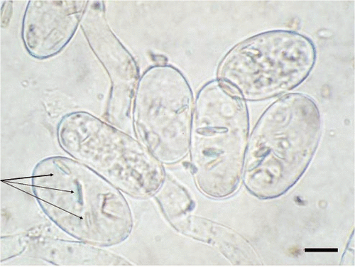
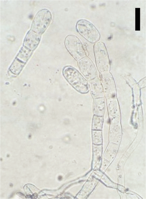
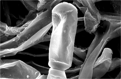
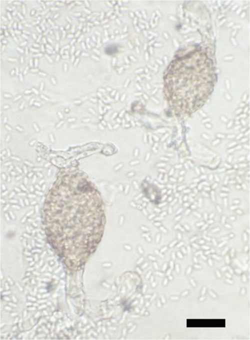
Previously, Golovinomyces, Podosphaera and Leveillula have been reported on Coreopsis (Amano 1986; Farr and Rossman 2009). Moreover, Oidium heliotropii-indici has been reported on Coreopsis from India (Paul and Thakur 2006). O. heliotropii-indici has nipple-shaped appressoria and this species is common on plants belonging to the family Boraginaceae. The specimens (AMH 9333, ICARHNEH-125) have ectophytic mycelium, conidia in chains, fibrosin bodies and indistinct appressoria, which indicated they belonged to the genus Podosphaera (Braun et al. 2002). Conidiophores were mostly erect, with some curved, and contained a foot cell (27–44 × 11–13.5 μm); conidia were ovoid, (27.5) 29–36 (–42) × (14.5–) 16–19 μm, with length : width 1.5–2.5 (av. 2) (Figs 2, 3). The basal septum of the conidiophore was just adjacent to the mycelium. Smooth wrinkles and indistinct appressoria (Fig. 4) were observed in SEM studies (Cook et al. 1997). Germination pattern was of Fibroidium subtype brevitubus, which is further evidence that this pathogen belonged to Podosphaera (section Sphaerotheca subsection Magnicellulatae) (Cook and Braun 2009). A perfect stage (chasmothecium) was not found. Species delimitation in the subsection Magnicellulatae is still controversial and requires revision (Hirata et al. 2000; Braun et al. 2001; Ito and Takamatsu 2010). Pathogenicity tests yielded positive results. Inoculated plants developed symptoms after 10 days, whereas control plants remained healthy. The hyperparasite Ampelomyces quisqualis was found associated with this pathogen (Fig. 5).
|
|
|
|
To our knowledge, this is the first record of powdery mildew of Coreopsis caused by Podosphaera sp. in India (Paul and Thakur 2006; Ahmed et al. 2007; Farr and Rossman 2009).
Acknowledgements
The authors would like to thank the Head, Sophisticated Analytical Instrument Facility, Dr Sudeep Dey (Scientific Officer), Dr R. Charkraborty, Mr. N. K. Rynjah for scanning electron microscopy at North-eastern Hill University, Shillong, Meghalaya, India.
Braun U,
Shishkoff N, Takamatsu S
(2001) Phylogeny of Podosphaera sect. Sphaerotheca subsect. Magnicellulatae (Sphaerotheca fuliginea auct. s. lat.) inferred from rDNA ITS sequences—a taxonomic interpretation. Schlechtendalia 7, 45–52.
(Last accessed 18 August 2010)
Hirata T,
Cunnington JH,
Paksiri U,
Limkaisang S,
Shishkoff N,
Grigaliunaite B,
Sato Y, Takamatsu S
(2000) Evolutionary analysis of subsection Magnicellulatae of Podosphaera section Sphaerotheca (Erysiphales) based on the rDNA ITS sequences with special reference to host plants. Canadian Journal of Botany 78, 1521–1530.
| Crossref | GoogleScholarGoogle Scholar |
CAS |

Ito M, Takamatsu S
(2010) Molecular phylogeny and evolution of subsection Magnicellulatae (Erysiphaceae: Podosphaera) with special reference to host plants. Mycoscience 51, 34–43.
| Crossref | GoogleScholarGoogle Scholar |
CAS |


