

Betulonic acid regulates oviduct epithelial cell inflammation through the TLR4, MAPK, and JAK/STAT signalling pathways
Liang Shao A , Yan Yan B , Nansu Wang B , Qiongfang Tan B , Yuying Huang B , Lei Lei C , Dongmei Yang D and Ling Liu B *
B *
A Department of Emergency, The First Affiliated Hospital of Hunan Traditional Chinese Medicine College, Zhuzhou, Hunan, China.
B Department of TCM Gynaecology, The First Affiliated Hospital of Hunan Traditional Chinese Medicine College, Zhuzhou, Hunan, China.
C Combine Traditional Chinese and Western Medicine Institute, Hunan University of Chinese Medicine, Changsha, Hunan, China.
D Medical School, Hunan University of Chinese Medicine, Changsha, Hunan, China.
Reproduction, Fertility and Development 35(8) 480-491 https://doi.org/10.1071/RD21380
Published online: 5 May 2023
Abstract
Infertility is a common disease among women of childbearing age and seriously endangers the reproductive health of human beings.
We aimed to study the active effect and mechanism of betulonic acid (BTA) on tubal inflammatory infertility.
An inflammatory model was established in isolated rat oviduct epithelial cells. Immunofluorescence of cytokeratin 18 was performed in cells. The therapeutic effect of BTA on cells was observed. Subsequently, we added JAK/STAT inhibitor AG490 and MAPK inhibitor U0126 and measured the levels of inflammatory factors via enzyme-linked immunosorbent assay and qRT-PCR. CCK-8 assay was applied to test cell proliferation, whereas flow cytometry was used to measure apoptosis. The levels of TLR4, IκBα, JAK1, JAK2, JAK3, Tyk2, STAT3, p38, ERK and the phosphorylation of p65 were determined by Western blotting.
Betulonic acid inhibited the activation of TLR4 and NF-κB signalling pathways, and significantly downregulated IL-1β, IL-6, and TNF-α, with high doses being the most effective. Furthermore, high-dose BTA promoted the proliferation of oviduct epithelial cells and inhibited apoptosis. In addition, BTA inhibited the activation of JAK/STAT signalling pathway to perform effectively in oviduct epithelial cells inflammation. The addition of AG490 led to the inhibition of the JAK/STAT signalling pathway. BTA also inhibited the activation of MAPK signalling pathway in oviduct epithelial cells inflammation. Under U0126 treatment, the inhibition of proteins in MAPK pathway by BTA was weakened.
Therefore, BTA inhibited the TLR, JAK/STAT and MAPK signalling pathways.
Our study provided a new therapeutic strategy for infertility caused by oviduct inflammation.
Keywords: betulonic acid, JAK, MAPK, NF-κB, rat oviduct epithelial cells, STAT, tubal inflammatory infertility, TLR4.
Introduction
Infertility is a common disease among women of childbearing age and seriously endangers the reproductive health of human beings (Vander Borght and Wyns 2018). Anatomical and functional causative factors are implicated in this condition. Structural abnormalities in the human anatomy, such as severe endometriosis or ectopic pregnancy and pelvic inflammation followed by tubal or uterine adhesions, are among the anatomical factors that result in infertility. Conversely, the abnormal function of reproductive organs caused by diseases such as autoimmune diseases, pelvic inflammatory disease, and salpingitis also leads to infertility (Yang 2016). Salpingitis is a part of pelvic inflammation, which is a threat for women’s health, accounts for 30–35% of female infertility (Zou et al. 2014; Yao et al. 2020). The oviduct is the site of oocyte and sperm transport, fertilisation, and early embryonic development. Infection and inflammation of the oviduct, which are the main causes of infertility in humans (Baczynska et al. 2007), are mainly caused by chlamydia, Neisseria gonorrhoeae, Escherichia coli, and other pathogens; repeated infection of the oviduct causes mucosa damage. Repeated attacks of local inflammation lead to salpingitis exudation, edema, and hyperplasia. The oviduct cavity gradually develops adhesion and stenosis, eventually causing oviduct obstructive infertility (Mardh 2004). Currently, laparoscopy and antibiotics are the dominating treatments for tubal factor infertility. However, postoperative obstruction of tubal repair caused by adhesions can lead to the recurrence of infertility. Moreover, the long-term use of antibiotics has side effects, which could lead to antibiotic abuse and bacterial resistance. Therefore, to manage salpingitis and the resulting oviduct obstruction, new treatment strategies are needed that can repair the physiology of the oviduct and reduce the infertility associated with salpingitis.
Increased expression of the inflammatory cytokines TNF-α, IL-1β, and TGF-β can aggravate the development of cell inflammation (Zhang et al. 2018). The activation of cellular signalling pathways includes the phosphorylation of the MAPK pathway and of specific transcription factors, i.e. NF-κB in combination with the cell-surface receptor Toll-like receptor 4 (TLR4) (Ren et al. 2020). The activation of TLR4, which is an important transmembrane pattern recognition receptor of the innate immune system, has been demonstrated in a variety of inflammatory conditions of the body. TLR4 is also an important gatekeeper for the innate immune system, as it recognises parts of the structure of certain conserved pathogens and continuously activates macrophages to produce an immune response. TLR4 is a receptor for chlamydia lipopolysaccharides (Prebeck et al. 2003) and heat-shock proteins (Bulut et al. 2002), and is normally stimulated by active chlamydia infection (Darville et al. 2003). There is also evidence that TLR4 plays a role in receptiveness to pathogens causing genital tract infections (Laisk et al. 2010). Specifically, the activation of TLR4 is associated with the expression of proinflammatory cytokines and activation of the NF-κB signalling pathway (Wu et al. 2015). Reportedly, the activation of the MAPK pathway, particularly the phosphorylation of ERK1/2, is increased during inflammation, which is important for its development. Once p-ERK is upregulated, p-ERK phosphorylates P65, which results in the activation of the downstream NF-κB pathway and the release of factors involved in different cellular processes (Meng et al. 2018). Increasing evidence suggests that continued JAK/STAT signalling pathway activation is closely associated with many immune and inflammatory diseases (Xin et al. 2020). Therefore, whether the TLR4, JAK/STAT, and MAPK pathways play an important role in oviduct inflammation warrants further study.
Traditional Chinese medicine (TCM) has been widely involved in the treatment of infertility (Liao et al. 2018; Xin et al. 2019). From the perspective of TCM, partial or complete obstruction of the oviduct is caused by qi stagnation and blood stasis in the lower abdomen. The main treatment is the removal of blood stasis, the dredging of collaterals, and the regulation of qi and blood (Xu et al. 2003). The Xuefu Zhuyu decoction is a famous compound in Chinese medicine recorded in the Yi Lin Gai Cuo since the Qing dynasty (1830 AD). This decoction contains 11 Chinese herbs (15 g of Angelica sinensis, 15 g of Rehmanniae radix, 15 g of Persicae semen, 10 g of Carthami flos, 10 g of Aurantii fructus, 10 g of Paeoniae radix rubra, 10 g of Chuanxiong rhizoma, 10 g of Bupleuri radix, 15 g of Platycodi radix, 10 g of Achyranthis bidentatae radix, and 5 g of Glycyrrhizae radix) and has anti-inflammatory, anti-oxidant, and angiogenic effects (Song et al. 2012; Xing et al. 2016). The Xuefu Zhuyu decoction is mostly used for promoting blood circulation and removing blood stasis (Dai et al. 2017).
In this study, we used the LC-MS method to develop the Oviduct Futong decoction based on the Xuefu Zhuyu decoction (15 g of Angelica sinensis, 15 g of Rehmanniae radix, 15 g of Persicae semen, 10 g of Carthami flos, 10 g of Aurantii fructus, 10 g of Paeoniae radix rubra, 10 g of Chuanxiong rhizoma, 6 g of Glycyrrhizae radix, 9 g of Manis squama, 15 g of Liquidambaris fructus, 10 g of Luffae fructus retinervus, 10 g of Gleditsiae spina, 10 g of Toosendan fructus, and 15 g of Spatholobi caulis), to add the function of collateral dredging by promoting blood circulation and removing blood stasis. Betulonic acid (BTA) was selected as the main component of the Oviduct Futong decoction. Subsequently, we performed several experiments to investigate the mechanism underlying the regulation of tubal inflammatory infertility afforded by BTA through the NF-κB, MAPK, and JAK/STAT signalling pathways.
Materials and methods
Isolation and identification of rat oviduct epithelial cells
The fallopian tubes of female SPF s.d. rats (sexually mature but had not copulated and had not given birth; at the oestrus stage of the oestrous cycle) were selected, and the rats were killed. The oviduct was collected and placed in 5 mL of Hank’s balanced salt solution (HBSS), and the fat and ovaries were removed from the tissues under an anatomical microscope. The oviduct was washed with HBSS. Tissues were cut into mincemeat form using ophthalmic scissors in Diss. The tissues were transferred to a culture bottle containing 0.25% trypsin and enzymatically hydrolysed for 30 min in a water bath shaker at 37°C. The mixture was transferred to a centrifuge tube and centrifuged at 201g for 5 min. After the supernatant was discarded, the tissue was transferred to a flask containing 1 mg/mL collagenase (HBSS) and enzymatically hydrolysed for 30 min at 37°C. The mixture was then transferred to a centrifuge tube and centrifuged at 201g for 5 min. The supernatant was discarded and 1 mL of DMEM/F-12 medium was added, to prepare a cell suspension, which was transferred to a disposable culture bottle. The superstratum cell suspension was sucked out in an incubator of 5% CO2 at 37°C for about 4 h, then transferred into a centrifuge tube and centrifuged at 201g for 5 min. The supernatant was discarded and 1 mL of DMEM/F-12 medium was added, followed by inoculation in a disposable culture dish after gently blowing and beating. The medium was cultured in a 5% CO2 incubator at 37°C for 48 h. Cell passage was performed when the oviduct epithelial cells exhibited 80% confluence or filled the plate floor. Immunofluorescence staining was used to identify the oviduct epithelial cells, and the positive rate of cytokeratin 18 was determined, to confirm that the cells were oviduct epithelial cells. After identification using cytokeratin 18, the three-passage cells were used for subsequent experiments.
Immunofluorescence
The positive rate of cytokeratin 18 was detected by immunofluorescence staining. The slices were removed and cleaned with PBS 2–3 times, then fixed with 4% paraformaldehyde for 30 min and rinsed with PBS 3 times (5 min each time). Subsequently, 0.5% TritonX-100 was used for permeabilisation for 30 min at 37°C. After washing with PBS, 5% BSA was closed at 37°C for 1 h, followed by incubation with an anti-cytokeratin 18 primary antibody at 4°C overnight, washing with PBS 3 times (5 min each time), incubation with a diluted fluorescent secondary antibody (Coralite 594-conjugated goat anti-rabbit IgG; SA00013-4, 1:200, Proteintech) at 37°C for 90 min, and rinsing with PBS 3 times (5 min each). DAPI working solution (Wellbio) was used to stain nuclei at 37°C for 10 min, and then rinsed with PBS 3 times (5 min each). The tablet was buffered with glycerin and observed under a fluorescence microscope.
Liquid chromatography/mass spectrometry (LC-MS)
The Oviduct Futong decoction solution (100 μL) was mixed with 400 μL of methanol–acetonitrile solution. The mixed solution was sonicated at 4°C for 10 min and stored at −20°C for 1 h. The mixture was centrifuged, and the precipitate was discarded. The supernatant was condensed into a dry powder via centrifugal freezing. The dry powder was dissolved in 50% acetonitrile and detected by LC-MS under the positive and negative modes. The instrument platform used for LC-MS analysis consisted of an Agilent 1290 ultra-performance liquid chromatographer and Thermo Fisher Scientific’s Q Precision Orbital RAP high-resolution mass spectrometer. The experiment was performed on an UPLC HSS T3 column (1.7 μm; 2.1 × 100 mm; Waters). A Q ExActive Orbitrap high-resolution mass spectrometer was used to collect the primary and secondary mass spectrometry data. The Compound Discoverer (ver. 2.0, Thermo Fisher) and OSI-SMMS (ver. 1.0, Dashuo) software were employed for material identification in combination with the MZCloud database and a self-built database. Finally, the normalised data matrix was used for multivariate statistical analysis, to screen the differential ingredients.
Establishment of an inflammatory model of rat oviduct epithelial cells
The rat oviduct epithelial cells were stimulated with LPS at different concentrations (1, 10, 50, 100, and 200 ng/mL), and the medium was used to replace LPS in the negative control (0 ng/mL LPS). Samples were collected at 0, 1, 3, 6, 12, and 24 h. The best dose and effect time of LPS were determined via the detection of IL-1β and other inflammatory factors. After determining the optimal dose and response time, the oviduct epithelial cells were divided into the control group (no LPS, no drug), the LPS group (model group, LPS-induced inflammation, no drug), the low-dose BTA group (low-BTA group, LPS-induced inflammation + 5 μM BTA), the medium-dose BTA group (med-BTA group, LPS-induced inflammation + 10 μM BTA), the high-dose BTA group (high-BTA group, LPS-induced inflammation + 20 μM BTA) (Dubinin et al. 2020), the JAK/STAT inhibitor group (LPS-induced inflammation + 10 μM AG490) (Merhi et al. 2013; Jiang et al. 2021), and the MAPK inhibitor group (LPS-induced inflammation + 10 μM U0126) (Yuan et al. 2021).
Enzyme-linked immunosorbent assay (ELISA)
Interleukin (IL)-1β, IL-6, and TNF-α were examined using the Quantitative ELISA Kit for IL-1β (#CSB-E08053h, C23037037, Cusabio), IL-6 (#CSB-E04638h, E31033828, Cusabio), and TNF-α (#CSB-E04740h, P05031137, Cusabio), according to the manufacturer’s instructions. The concentrations of IL-1β, IL-6, and TNF-α in cells were calculated using the standard curve provided by the DYNATECHMR7000 microplate analyser, and the results were expressed as pg/mL.
Quantitative real-time PCR (qRT-PCR)
Total RNA was extracted using the Trizol method. The cDNA of PrimeScript II was synthesised by Takara. mRNA expression was detected by qRT-PCR using the SYBR GREEN PC Master Mix (Applied Biosystems). The reaction was carried out on an ABI PRISM 7300 sequence detection system (ASB). The relative quantitative equation 2−ΔΔCt was used to calculate mRNA expression levels. β-actin served as an internal reference. The primers used here were as follows: IL-1β-F: TGTGATGTTCCCATTAGAC, IL-1β-R: AATACCACTTGTTGGCTTA; IL-6-F: TCACTATGAGGTCTACTCGG, IL-6-R: CATATTGCCAGTTCTTCGTA; TNF-α-F: CCCCTCTATTTATAATTGCACCT, TNF-α-R: CTGGTAGTTTAGCTCCGTTT; and β-actin-F: ACATCCGTAAAGACCTCTATGCC, β-actin-R: TACTCCTGCTTGCTGATCCAC.
Western blotting
RIPA buffer (#P0013B, Beyotime) was used to extract total proteins from cells according to the manufacturer’s instructions, and proteins were quantified using a BCA protein assay kit. The mixed SDS–PAGE loading buffer (#MB2479, Meilunbio) was heated in a water bath for 5 min at 100°C. The proteins were separated by gel electrophoresis and transferred to a PVDF membrane. The membrane was closed with a 5% skim milk solution for 90 min at room temperature, followed by incubation with antibodies against TLR4 (19 811-1-AP, 1: 750, Proteintech), p65 (10 745-1-AP, 1:2000, Proteintech), p-p65 (bs-0982r, 1:1000, Bioss), IκBα (10 268-1-AP, 1:2000, Proteintech), p-IκBα (9246s, 1:1000, CST), p38 (14 064-1-AP, 1:1500, Proteintech), p-p38 (ab4822, 1:1000, Abcam), ERK (11 257-1-AP, 1:1000, Proteintech), p-ERK (ab50011, 1:4000, Abcam), JNK (ab179461, 1:1000, Abcam), p-JNK (ab124956, 1:4000, Abcam), JAK1 (66 466-1-Ig, 1:2000, Proteintech), p-JAK1 (ab138005, 1:3000, Abcam), JAK2 (17 670-1-AP, 1:1000, Proteintech), p-JAK2 (ab32101, 1:3000, Abcam), JAK3 (ab203611, 1:750, Abcam), p-JAK3 (bs-20168 R, 1:1000, Bioss), Tyk2 (bs-6662 R, 1:1000, Bioss), p-Tyk2 (bs-3437 R, 1:1000, Bioss), STAT3 (10 253-2-AP, 1:3000, Proteintech), p-STAT3 (ab76315, 1:4000, Abcam), and GAPDH (10 494-1-AP, 1:4000, Proteintech) at 4°C overnight. Subsequently, the membranes were incubated with HRP-conjugated goat anti-mouse IgG (SA00001-1, 1:5000, Proteintech) and HRP-conjugated goat anti-Rabbit IgG (SA00001-2, 1:6000, Proteintech). Detection was performed using ECL, and an Odyssey Infrared Imaging System (Li-Cor Biosciences) was used to detect protein bands using GAPDH as the internal reference to determine expression levels.
Cell counting kit-8 (CCK-8) assay
The cells were digested, counted and inoculated in 96-well plates at a density of 1 × 10⁴ cells/well, 100 μL per well, followed by incubation at 37°C and 5% CO2. After treatment for 0, 12, 24, and 48 h, 10 μL of CCK-8 (#NU679, DOJINDO, Japan) was added to each well. The absorbance (OD) at 450 nm was determined on a bio-Tek microplate (MB-530, Heales, China) after further incubation at 37°C and 5% CO2 for 4 h.
Cell apoptosis
Cells were digested with trypsin without EDTA and collected. The cells were washed with PBS twice, centrifuged for 5 min at 402g each time, and about 5 × 10⁵ cells were collected. We added 500 μL of binding buffer to suspend the cells and 5 μL of Annexin V-FITC (KGA108, KeyGen, China), and the solution was mixed. Subsequently, we added 5 μL of propidium iodide and mixed the solution. The reaction was carried out for 15 min in the dark at room temperature. The results were observed and detected by flow cytometry (A00-1 1102, Beckman) within 1 h.
Results
Establishment of an inflammatory model of oviduct epithelial cells
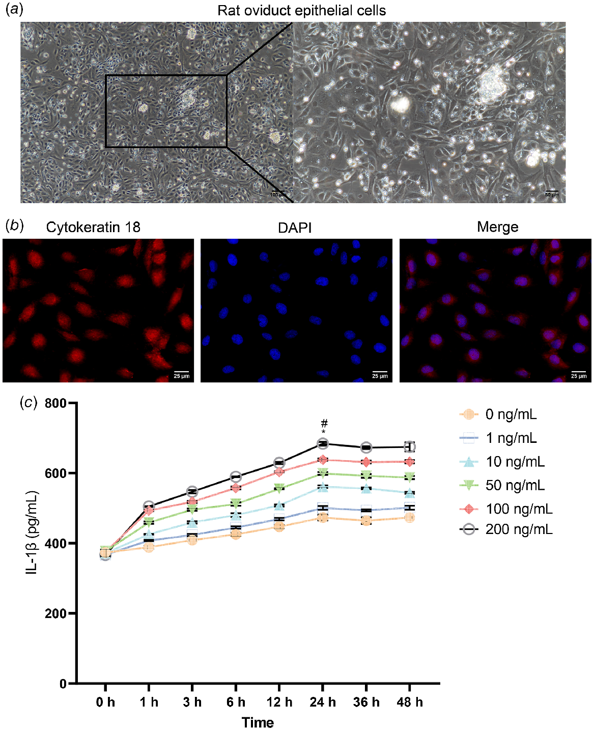
Oviduct epithelial cells were isolated from rats. Fig. 1a showed the image of the cultured oviduct epithelial cells. Immunofluorescence staining showed that cytokeratin 18 was present in the cells (Fig. 1b). These results confirmed that the isolated cells were oviduct epithelial cells. LPS was used to induce oviduct epithelial cells, to establish an inflammatory model (Fig. 1c). The effect of stimulation using different concentrations of LPS (0 ng/mL, 1 ng/mL, 10 ng/mL, 50 ng/mL, 100 ng/mL, and 200 ng/mL) on the epithelial cells was investigated. Samples were collected at 0, 1, 3, 6, 12, and 24 h. According to the level of the inflammatory factor IL-1β in the supernatant, the best dose and effect time of LPS were determined to be 200 ng/mL and 24 h, respectively.
Establishment of an inflammatory model of oviduct epithelial cells. (a) The images of the cultured oviduct epithelial cells (100×, 100 μm; 200×, 50 μm). (b) Immunofluorescence staining of cytokeratin 18 in oviduct epithelial cells (100×, 100 μm). (c) Changes in the concentration of IL-1β induced by different doses of LPS over time. *P < 0.05 vs 0 h, # P < 0.05 vs 0 ng/mL.
Inhibitory effect of BTA on the TLR4 pathway in oviduct inflammation
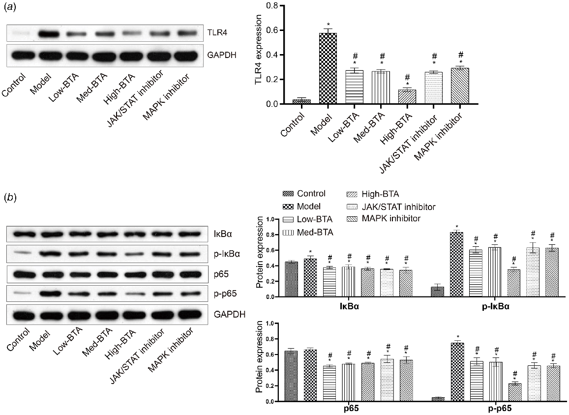
Liquid chromatography-MS was used to analyse the key components of the Oviduct Futong decoction under the positive and negative modes (Supplementary Fig. S1). Positive mode highlights the difference between protocols of mass spectra measurement, such as fresh and frozen–thawed samples, whereas negative mode better characterises the histological difference between samples (Zhvansky et al. 2021). Dual analysis in both positive and negative ion modes is a potential approach to identify peptides with unstable modifications, such as sulfation, and to distinguish them from phosphorylation (Drake and Hortin 2010). The data obtained in the two ion modes are complementary to some extent (Nicolardi et al. 2020). Comparison of positive and negative ion ESI-MS spectra might provide an insight into various binding modes in both solution and the gas phase (Gupta et al. 2004). Based on the value of area, by analysing the ingredients in Oviduct Futong decoction, we found that, under the positive mode, apigenin 7-O-beta-d-glucuronide, ethanolamine phosphate, BTA, patchouli alcohol and dibutyl phthalate were the main ingredients of the Oviduct Futong decoction. Under the negative mode, maltopentaose, furfuryl alcohol, fungitetraose, stachyose and mimosine were the main ingredients of the decoction. These ingredients might further contribute to the beneficial effect of the decoction on tubal inflammatory infertility (Supplementary Table S1). After reviewing the literature, we found that BTA has anti-inflammatory activity (Vasilevsky et al. 2009; Popov et al. 2019; Song et al. 2021; Semenova et al. 2022). In addition, BTA has rarely been studied in tubal inflammatory infertility. Therefore, we chose BTA to perform follow-up experiments. To explore the potential molecular mechanism underlying the beneficial effect of BTA on oviduct inflammation, we investigated the therapeutic effects of BTA on oviduct inflammatory cells and their roles in the TLR4/NF-κB receptor pathway. We monitored TLR4 expression in the cells. The results revealed that the levels of the TLR4 protein were significantly increased in the model group, whereas the TLR4 protein was downregulated in the BTA groups, with the TLR4 protein being more significantly downregulated in the high-BTA group (Fig. 2a). Interestingly, compared with the control group, the phosphorylation (activation) of p65 and IκBα in the model group was increased, whereas the phosphorylation (activation) of p65 and IκBα in all BTA groups was decreased, with the phosphorylation (activation) of p65 and IκBα in the high-BTA group being more significantly decreased (Fig. 2b). These results suggest that high-dose BTA inhibits the activation of the TLR4 and NF-κB signalling pathways.
High-dose BTA inhibited the inflammatory response, promoted oviduct epithelial cell proliferation, and inhibited apoptosis
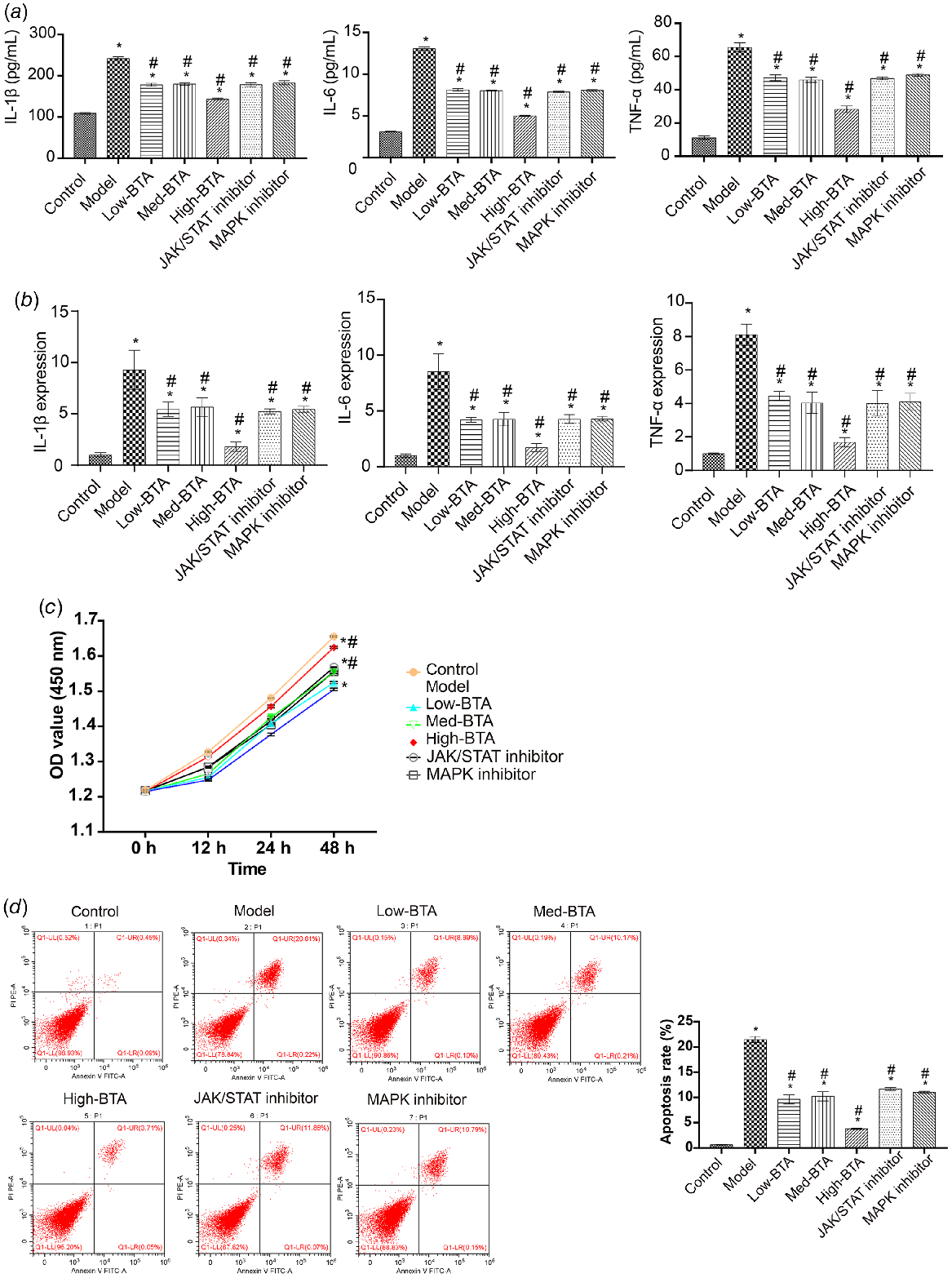
To assess further the effects of BTA, we investigated the therapeutic effects of BTA on oviduct inflammatory cells. First, we measured changes in inflammatory markers. Compared with the control group, IL-1β, IL-6, and TNF-α were upregulated in the model group. After adding BTA, the levels of IL-1β, IL-6, and TNF-α were decreased (Fig. 3a, b), with high-dose BTA having the best inhibitory effect on inflammation. Next, we measured changes in cellular function. We found decreased proliferation and increased apoptosis in the model group compared with the control group. After adding BTA, proliferation increased and apoptosis decreased, with the inhibitory effect of high-dose BTA being more significant (Fig. 3c, d). These results suggest that high-dose BTA inhibits the inflammatory response, promotes the proliferation of oviduct epithelial cells, and inhibits apoptosis.
High-dose BTA inhibited the inflammatory response, promoted oviduct epithelial cell proliferation, and inhibited apoptosis. (a) The levels of IL-1β, IL-6, and TNF-α were detected by ELISA. (b) qRT-PCR was used to assess IL-1β, IL-6, and TNF-α levels. (c) The CCK-8 assay was applied to detect cell proliferation in each group. (d) Flow cytometry was used to examine apoptosis in each group. *P < 0.05 vs the control, #P < 0.05 vs the model.
High-dose BTA inhibited the JAK/STAT signalling pathway
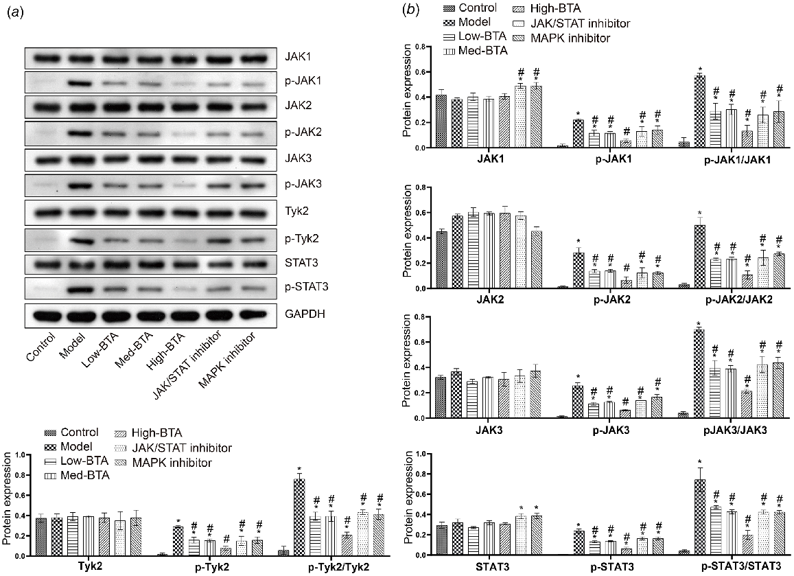
Subsequently, to confirm the effects of BTA on the JAK/STAT signalling pathway, we assessed the expression of JAK1, JAK2, JAK3, Tyk2 and STAT3. The results showed that the JAK/STAT signalling pathway was activated in oviduct inflammation, and that the phosphorylation levels of the JAK1, JAK2, JAK3, Tyk2 and STAT3 proteins were increased. Next, the activation of JAK1, JAK2, JAK3, Tyk2 and STAT3 was inhibited by BTA. After adding AG490, which is a JAK/STAT pathway inhibitor, the JAK/STAT pathway was inhibited (Fig. 4a, b). Based on these results, we conclude that BTA inhibits the activation of the JAK/STAT signalling pathway to perform effectively in oviduct epithelial cells inflammation.
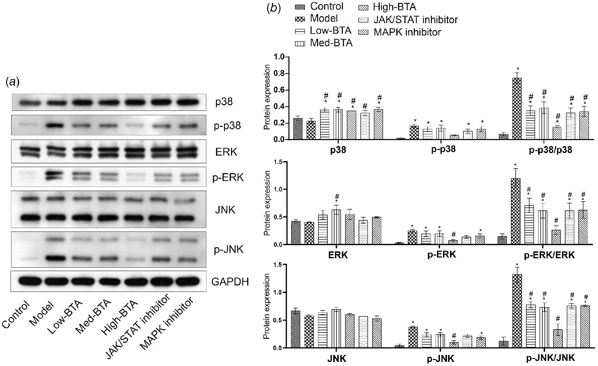
High-dose BTA inhibited the MAPK signalling pathway
Next, to confirm the effects of BTA on the MAPK signalling pathway, we measured the levels of p38, ERK, and JNK in an established oviduct inflammation model. As shown in Fig. 5a, b, the MAPK signalling pathway was activated in oviduct inflammation. In turn, the activation of the p38, ERK, and JNK proteins was inhibited by BTA. Under the action of U0126 (which is a MAPK pathway inhibitor), the MAPK signalling pathway was inhibited. Based on these results, we concluded that BTA inhibits the activation of the MAPK signalling pathway in oviduct epithelial cells inflammation.
Discussion
Chronic salpingitis is a significant cause of infertility in women. TCM has a variety of methods for the treatment of chronic salpingitis and has achieved relatively good clinical efficacy, which underscores the advantages of TCM (Lian et al. 1991; Kang et al. 2001; Wen et al. 2015). The main component of the Oviduct Futong decoction, BTA, has been shown to have anti-inflammatory activity (Vasilevsky et al. 2009; Popov et al. 2019). In this study, the oviduct epithelial cells isolated from the oviduct in vitro were used to show that BTA has an anti-inflammatory effect on oviduct epithelial cells, which significantly downregulates IL-1β, IL-6 and TNF-α. Moreover, the therapeutic effect of BTA was not only related to the TLR pathway, but also possessed anti-inflammatory activity by inhibiting the activation of the JAK/STAT and MAPK signalling pathways.
Innate immunity is triggered when the body is stimulated by xenobiotics, followed by the induction of TLR4 on the cell surface, to activate specific immunity (Jiang et al. 2020). A previous study showed that the immune response activates the NF-κB inflammatory pathway via the TLR4 receptor protein, leading to large releases of the proinflammatory cytokines TNF-α and IL-1β into the extracellular matrix (O’Neill and Bowie 2007). The increased production and release of proinflammatory factors further activates NF-κB, which results in the continuous amplification of the initial inflammatory signals, increased permeability of the epithelial cell layer, and tissue injury (Huang et al. 2016; Zhang et al. 2017). Similarly, a previous study has demonstrated that the activation of the TLR and NF-κB pathways by external infection leads to salpingitis (Shaw et al. 2011). As mentioned above, our results showed that the expression of IL-1β, IL-6 and TNF-α was significantly higher in the model group than it was in the control group. BTA effectively reduced the expression of proinflammatory factors in oviduct cells. Subsequently, we found that BTA reduced TLR4 expression and p65 and IκBα phosphorylation in inflammatory epithelial cells. Again, high-dose BTA had a superior therapeutic effect. These results suggest that the inhibitory effect of BTA on oviduct epithelial cells inflammation may be related to TLR4. In addition, the mechanism of oviduct epithelial cells inflammation by BTA requires further study.
The JAK/STAT pathway is a classic inflammatory pathway. Inhibition of this pathway inhibits α-synuclein-induced neuroinflammation and dopaminergic neurodegeneration (Qin et al. 2016). Cheng et al. (2020) reported that cinnamaldehyde, a bioactive cinnamon essential oil from the TCM herb Cinnamomum cassia, inhibited the inflammation of human synoviocyte cells by regulating the JAK/STAT pathway and ameliorating collagen-induced arthritis in rats. In our study, after adding AG490, which is a JAK/STAT pathway inhibitor, the JAK/STAT pathway was inhibited. In contrast, BTA inhibited the activation of the JAK/STAT signalling pathway to play a role in oviduct epithelial cells inflammation.
The MAPK family consists of at least three members, i.e. ERK, C-Jun N-terminal kinase (JNK), and p38 MAPK, which play a vital role in cell-cycle regulation, proliferation, and cell survival (Garrington and Johnson 1999; Kyriakis and Avruch 2012). The p38 mitogen-activated protein kinase (MAPK) is thought to be a key mediator of the activation of the inflammatory factors TNF-α, IL-1β, and IL-6 (Kraatz et al. 1999; Shames et al. 1999). MAPK and JNK were shown to be helpful in upregulating TNF-α and enhancing cell inflammation and apoptosis (Chen et al. 2018). The activation of ERK was mainly involved in the senescence and proliferation of inflammatory cells (Yang et al. 2013). This study confirmed that p38 MAPK, ERK, and JNK were activated after LPS-induced oviduct epithelial cell inflammation. However, BTA reversed the effects of LPS on the MAPK family of proteins in epithelial cells. Notably, our results demonstrated that U0126 significantly increased the phosphorylation of p38 MAPK, ERK, and JNK in the BTA groups. These results suggest that inhibition of MAPK activation might be the main mechanism via which BTA reduces cell inflammation.
However, in this study, using primary oviduct epithelial cell culture for experiments has limitations. Ciliated epithelial cells lost their cilia and beating function after culture and passages. Therefore, the results obtained are not applicable to all epithelial cells in the oviduct. Moreover, the findings in this study are applicable to a cell culture model and might not be applicable to these situations during pregnancy. In the future, some experiments could be validated in vivo through collection of whole oviducts after the LPS treatment with the BTA and performing ELISA or western blotting. In order to specifically observe changes in epithelial cells, we should collect oviductal sections for immunohistochemistry/immunofluorescence to show the presence of cytokines with or without BTA treatment. Moreover, we should perform experiments in pregnant females to conclude our findings were applicable to the situation during pregnancy. Furthermore, we didn’t verify whether BTA would be safe during pregnancy. We also didn’t know how BTA could affect embryo development. Therefore, we should perform fertility trial in BTA-treated pregnant rats, and collect the embryos to ensure that embryo development is not disrupted in the presence of BTA (e.g. quantify the percentage of blastocysts produced in BTA-treated and control groups). Alternatively, we should also culture zygotes with BTA to observe its effect on blastocyst development. However, due to time and funding constraints, we cannot yet solve them well. With sufficient time and funding in the future, we will conduct a series of related in vivo experiments.
In conclusion, our research demonstrated that BTA inhibited the TLR, JAK/STAT, and MAPK signalling pathways, suggesting that BTA could be a potential therapeutic strategy for treating infertility caused by oviduct inflammation in future researches. Moreover, our study has important significance for TCM theory and mechanistic research of TCM monomers.
Data availability
The data that support this study will be shared upon reasonable request to the corresponding author.
Declaration of funding
This work was supported by the National Natural Science Foundation of China (81804140), the China Postdoctoral Science Foundation (2017M612568), the Natural Science Foundation of Hunan Province (2018JJ3407), the Project of Hunan Province “Shennong Talents” for Young Shennong Scholars, the Project of Hunan Province “Hunan Young Talents”, the Research Project on Chinese Medicine of Hunan Province (A2023052) and the Natural Science Foundation of Zhuzhou.
References
Baczynska A, Funch P, Fedder J, Knudsen HJ, Birkelund S, Christiansen G (2007) Morphology of human fallopian tubes after infection with Mycoplasma genitalium and Mycoplasma hominis—in vitro organ culture study. Human Reproduction 22(4), 968-979.
| Crossref | Google Scholar |
Bulut Y, Faure E, Thomas L, Karahashi H, Michelsen KS, Equils O, Morrison SG, Morrison RP, Arditi M (2002) Chlamydial heat shock protein 60 activates macrophages and endothelial cells through toll-like receptor 4 and MD2 in a MyD88-dependent pathway. Journal of Immunology 168(3), 1435-1440.
| Crossref | Google Scholar |
Chen H, Wang X, Yan X, Cheng X, He X, Zheng W (2018) RETRACTED: LncRNA MALAT1 regulates sepsis-induced cardiac inflammation and dysfunction via interaction with miR-125b and p38 MAPK/NFκB. International Immunopharmacology 55, 69-76.
| Crossref | Google Scholar |
Cheng WX, Zhong S, Meng XB, Zheng NY, Zhang P, Wang Y, Qin L, Wang XL (2020) Cinnamaldehyde inhibits inflammation of human synoviocyte cells through regulation of JAK/STAT pathway and ameliorates collagen-induced arthritis in rats. Journal of Pharmacology and Experimental Therapeutics 373(2), 302-310.
| Crossref | Google Scholar |
Dai L, Lu A, Zhong LLD, Zheng G, Bian Z (2017) Chinese herbal medicine for hyperlipidaemia: a review based on data mining from 1990 to 2016. Current Vascular Pharmacology 15(6), 520-531.
| Crossref | Google Scholar |
Darville T, O’Neill JM, Andrews CW, Jr, Nagarajan UM, Stahl L, Ojcius DM (2003) Toll-like receptor-2, but not toll-like receptor-4, is essential for development of oviduct pathology in chlamydial genital tract infection. Journal of Immunology 171(11), 6187-6197.
| Crossref | Google Scholar |
Drake SK, Hortin GL (2010) Improved detection of intact tyrosine sulfate-containing peptides by matrix-assisted laser desorption/ionization time-of-flight mass spectrometry in linear negative ion mode. International Journal of Biochemistry & Cell Biology 42(1), 174-179.
| Crossref | Google Scholar |
Dubinin MV, Semenova AA, Ilzorkina AI, Mikheeva IB, Yashin VA, Penkov NV, Vydrina VA, Ishmuratov GY, Sharapov VA, Khoroshavina EI, Gudkov SV, Belosludtsev KN (2020) Effect of betulin and betulonic acid on isolated rat liver mitochondria and liposomes. Biochimica et Biophysica Acta (BBA) - Biomembranes 1862(10), 183383.
| Crossref | Google Scholar |
Garrington TP, Johnson GL (1999) Organization and regulation of mitogen-activated protein kinase signaling pathways. Current Opinion in Cell Biology 11(2), 211-218.
| Crossref | Google Scholar |
Gupta R, Beck JL, Ralph SF, Sheil MM, Aldrich-Wright JR (2004) Comparison of the binding stoichiometries of positively charged DNA-binding drugs using positive and negative ion electrospray ionization mass spectrometry. Journal of the American Society for Mass Spectrometry 15(10), 1382-1391.
| Crossref | Google Scholar |
Huang Z, Zhuang X, Xie C, Hu X, Dong X, Guo Y, Li S, Liao X (2016) Exogenous hydrogen sulfide attenuates high glucose-induced cardiotoxicity by inhibiting NLRP3 inflammasome activation by suppressing TLR4/NF-κB pathway in H9c2 cells. Cellular Physiology and Biochemistry 40(6), 1578-1590.
| Crossref | Google Scholar |
Jiang G, Gong M, Song H, Sun W, Zhao W, Wang L (2020) Tob2 inhibits TLR-induced inflammatory responses by association with TRAF6 and MyD88. Journal of Immunology 205(4), 981-986.
| Crossref | Google Scholar |
Jiang S, Wang H, Zhou Q, Li Q, Liu N, Li Z, Chen C, Deng Y (2021) Melatonin ameliorates axonal hypomyelination of periventricular white matter by transforming A1 to A2 astrocyte via JAK2/STAT3 pathway in septic neonatal rats. Journal of Inflammation Research 14, 5919-5937.
| Crossref | Google Scholar |
Kang JL, Xia W, He QY (2001) Clinical study on treatment of oviduct obstruction by integrative traditional Chinese and Western Medicine. Zhongguo Zhong Xi Yi Jie He Za Zhi 21(6), 416-418 [In Chinese].
| Google Scholar |
Kraatz J, Clair L, Rodriguez JL, West MA (1999) Macrophage TNF secretion in endotoxin tolerance: role of SAPK, p38, and MAPK. Journal of Surgical Research 83(2), 158-164.
| Crossref | Google Scholar |
Kyriakis JM, Avruch J (2012) Mammalian MAPK signal transduction pathways activated by stress and inflammation: a 10-year update. Physiological Reviews 92(2), 689-737.
| Crossref | Google Scholar |
Laisk T, Peters M, Saare M, Haller-Kikkatalo K, Karro H, Salumets A (2010) Association of CCR5, TLR2, TLR4 and MBL genetic variations with genital tract infections and tubal factor infertility. Journal of Reproductive Immunology 87(1–2), 74-81.
| Crossref | Google Scholar |
Lian F, Sun NQ, Xia GC (1991) Experimental and clinical study of tong jing bao and angelicae complex injection in treating fallopian tube obstruction. Zhong Xi Yi Jie He Za Zhi 11(5), 282-285 [In Chinese].
| Google Scholar |
Liao WT, Chiang JH, Li CJ, Lee MT, Su CC, Yen HR (2018) Investigation on the use of traditional Chinese medicine for polycystic ovary syndrome in a nationwide prescription database in Taiwan. Journal of Clinical Medicine 7(7), 179.
| Crossref | Google Scholar |
Mardh P-A (2004) Tubal factor infertility, with special regard to chlamydial salpingitis. Current Opinion in Infectious Diseases 17(1), 49-52.
| Crossref | Google Scholar |
Meng LL, Wang JL, Xu SP, Zu LD, Yan ZW, Zhang JB, Han YQ, Fu GH (2018) Low serum gastrin associated with ER+ breast cancer development via inactivation of CCKBR/ERK/P65 signaling. BMC Cancer 18(1), 824.
| Crossref | Google Scholar |
Merhi Z, Buyuk E, Berger DS, Zapantis A, Israel DD, Chua S, Jr, Jindal S (2013) Leptin suppresses anti-Mullerian hormone gene expression through the JAK2/STAT3 pathway in luteinized granulosa cells of women undergoing IVF. Human Reproduction 28(6), 1661-1619.
| Crossref | Google Scholar |
Nicolardi S, Kilgour DPA, van der Burgt YEM, Wuhrer M (2020) Improved N- and C-terminal sequencing of proteins by combining positive and negative ion MALDI in-source decay mass spectrometry. Analytical Chemistry 92(18), 12429-12436.
| Crossref | Google Scholar |
O’Neill LAJ, Bowie AG (2007) The family of five: TIR-domain-containing adaptors in toll-like receptor signalling. Nature Reviews Immunology 7(5), 353-364.
| Crossref | Google Scholar |
Popov SA, Semenova MD, Baev DS, Sorokina IV, Zhukova NA, Frolova TS, Tolstikova TG, Shults EE, Turks M (2019) Lupane-type conjugates with aminoacids, 1,3,4-oxadiazole and 1,2,5-oxadiazole-2-oxide derivatives: Synthesis, anti-inflammatory activity and in silico evaluation of target affinity. Steroids 150, 108443.
| Crossref | Google Scholar |
Prebeck S, Brade H, Kirschning CJ, da Costa CP, Dürr S, Wagner H, Miethke T (2003) The gram-negative bacterium chlamydia trachomatis L2 stimulates tumor necrosis factor secretion by innate immune cells independently of its endotoxin. Microbes and Infection 5(6), 463-470.
| Crossref | Google Scholar |
Qin H, Buckley JA, Li X, Liu Y, Fox TH, 3rd, Meares GP, Yu H, Yan Z, Harms AS, Li Y, Standaert DG, Benveniste EN (2016) Inhibition of the JAK/STAT pathway protects against α-synuclein-induced neuroinflammation and dopaminergic neurodegeneration. Journal of Neuroscience 36(18), 5144-5159.
| Crossref | Google Scholar |
Ren Q, Guo F, Tao S, Huang R, Ma L, Fu P (2020) Flavonoid fisetin alleviates kidney inflammation and apoptosis via inhibiting Src-mediated NF-κB p65 and MAPK signaling pathways in septic AKI mice. Biomedicine & Pharmacotherapy 122, 109772.
| Crossref | Google Scholar |
Semenova MD, Popov SA, Sorokina IV, Meshkova YV, Baev DS, Tolstikova TG, Shults EE (2022) Conjugates of lupane triterpenoids with arylpyrimidines: synthesis and anti-inflammatory activity. Steroids 184, 109042.
| Crossref | Google Scholar |
Shames BD, Selzman CH, Pulido EJ, Meng X, Meldrum DR, McIntyre RC, Jr., Harken AH, Banerjee A (1999) LPS-induced NF-κB activation and TNF-α release in human monocytes are protein tyrosine kinase dependent and protein kinase C independent. Journal of Surgical Research 83(1), 69-74.
| Crossref | Google Scholar |
Shaw JLV, Wills GS, Lee KF, Horner PJ, McClure MO, Abrahams VM, Wheelhouse N, Jabbour HN, Critchley HOD, Entrican G, Horne AW (2011) Chlamydia trachomatis infection increases fallopian tube PROKR2 via TLR2 and NFκB activation resulting in a microenvironment predisposed to ectopic pregnancy. The American Journal of Pathology 178(1), 253-260.
| Crossref | Google Scholar |
Song J, Chen WY, Wu LY, Zheng LP, Lin W, Gao D, Kaptchuk TJ, Chen KJ (2012) A microarray analysis of angiogenesis modulation effect of Xuefu Zhuyu decoction on endothelial cells. Chinese Journal of Integrative Medicine 18(7), 502-506.
| Crossref | Google Scholar |
Song KN, Lu YJ, Chu CJ, Wu YN, Huang HL, Fan BY, Chen GT (2021) Biotransformation of betulonic acid by the fungus Rhizopus arrhizus CGMCC 3.868 and antineuroinflammatory activity of the biotransformation products. Journal of Natural Products 84(10), 2664-2674.
| Crossref | Google Scholar |
Vander Borght M, Wyns C (2018) Fertility and infertility: definition and epidemiology. Clinical Biochemistry 62, 2-10.
| Crossref | Google Scholar |
Vasilevsky SF, Govdi AI, Shults EE, Shakirov MM, Sorokina IV, Tolstikova TG, Baev DS, Tolstikov GA, Alabugin IV (2009) Efficient synthesis of the first betulonic acid-acetylene hybrids and their hepatoprotective and anti-inflammatory activity. Bioorganic and Medicinal Chemistry 17(14), 5164-5169.
| Crossref | Google Scholar |
Wen H, Fu J, Tang H, Ge M, Feng L (2015) Hydrotubation combined with Chinese herbal medicine for salpingitic infecundity: a systematic review and meta-analysis. Cell Biochemistry and Biophysics 71(2), 519-527.
| Crossref | Google Scholar |
Wu J, Li L, Sun Y, Huang S, Tang J, Yu P, Wang G (2015) Altered molecular expression of the TLR4/NF-κB signaling pathway in mammary tissue of Chinese holstein cattle with mastitis. PLoS ONE 10(2), e0118458.
| Crossref | Google Scholar |
Xin M, He J, Zhang Y, Wu Y, Yang W, Liang X, Yin X (2019) Chinese herbal decoction of Wenshen Yangxue formula improved fertility and pregnancy rate in mice through PI3K/Akt signaling. Journal of Cellular Biochemistry 120(3), 3082-3090.
| Crossref | Google Scholar |
Xin P, Xu X, Deng C, Liu S, Wang Y, Zhou X, Ma H, Wei D, Sun S (2020) The role of JAK/STAT signaling pathway and its inhibitors in diseases. International Immunopharmacology 80, 106210.
| Crossref | Google Scholar |
Xing Z, Xia Z, Peng W, Li J, Zhang C, Fu C, Tang T, Luo J, Zou Y, Fan R, Liu W, Xiong X, Huang W, Sheng C, Gan P, Wang Y (2016) Xuefu Zhuyu decoction, a traditional Chinese medicine, provides neuroprotection in a rat model of traumatic brain injury via an anti-inflammatory pathway. Scientific Reports 6, 20040.
| Crossref | Google Scholar |
Xu X, Yin H, Tang D, Zhang L, Gosden RG (2003) Application of traditional Chinese medicine in the treatment of infertility. Human Fertility 6(4), 161-168.
| Crossref | Google Scholar |
Yang XJ (2016) Telocytes in inflammatory gynaecologic diseases and infertility. Advances in Experimental Medicine and Biology 913, 263-285.
| Crossref | Google Scholar |
Yang P, Han Y, Gui L, Sun J, Chen YL, Song R, Guo JZ, Xie YN, Lu D, Sun L (2013) Gastrodin attenuation of the inflammatory response in H9c2 cardiomyocytes involves inhibition of NF-κB and MAPKs activation via the phosphatidylinositol 3-kinase signaling. Biochemical Pharmacology 85(8), 1124-1133.
| Crossref | Google Scholar |
Yao L, Xu B, Li X (2020) Neisseria gonorrhoeae-induced salpingitis is targeted by circular RNA EIF3K via miR-139-5p and regulating MAPK/NF-κB signaling pathway to promotes apoptosis and autophagy bacterial cells. Microbial Pathogenesis 142, 104051.
| Crossref | Google Scholar |
Yuan J, Liang X, Zhou W, Feng J, Wang Z, Shen S, Guan X, Zhao L, Deng F (2021) TRPA1 promotes cisplatin-induced nephrotoxicity through inflammation mediated by the MAPK/NF-κB signaling pathway. Annals of Translational Medicine 9(20), 1578.
| Crossref | Google Scholar |
Zhang X, Du Q, Yang Y, Wang J, Dou S, Liu C, Duan J (2017) The protective effect of luteolin on myocardial ischemia/reperfusion (I/R) injury through TLR4/NF-κB/NLRP3 inflammasome pathway. Biomedicine & Pharmacotherapy 91, 1042-1052.
| Crossref | Google Scholar |
Zhang J, Yu C, Zhang X, Chen H, Dong J, Lu W, Song Z, Zhou W (2018) Porphyromonas gingivalis lipopolysaccharide induces cognitive dysfunction, mediated by neuronal inflammation via activation of the TLR4 signaling pathway in C57BL/6 mice. Journal of Neuroinflammation 15(1), 37.
| Crossref | Google Scholar |
Zhvansky ES, Eliferov VA, Sorokin AA, Shurkhay VA, Pekov SI, Bormotov DS, Ivanov DG, Zavorotnyuk DS, Bocharov KV, Khaliullin IG, Belenikin MS, Potapov AA, Nikolaev EN, Popov IA (2021) Assessment of variation of inline cartridge extraction mass spectra. Journal of Mass Spectrometry 56(4), e4640.
| Crossref | Google Scholar |
Zou SE, Jin Y, Ko YL, Zhu J (2014) A new classification system for pregnancy prognosis of tubal factor infertility. International Journal of Clinical and Experimental Medicine 7(5), 1410-1416.
| Google Scholar |