

Prenatal dexamethasone exposure and developmental programming of the ovary of the offspring: a structural study in the rat
Nataša Ristić A C , Nataša Nestorović A , Milica Manojlović-Stojanoski A , Svetlana Trifunović A , Vladimir Ajdžanović A , Branko Filipović A , Lazo Pendovski B and Verica Milošević A
A Institute for Biological Research ‘Siniša Stanković’, National Institute of Republic of Serbia, University of Belgrade, Bulevar despota Stefana 142, 11060 Belgrade, Serbia.
B Faculty of Veterinary Medicine, Ss. Cyril and Methodius University in Skopje, Lazar Pop-Trajkov 5-7 1000 Skopje, Republic of North Macedonia.
C Corresponding author. Email: negicn@ibiss.bg.ac.rs
Reproduction, Fertility and Development 33(3) 245-255 https://doi.org/10.1071/RD20164
Submitted: 22 June 2020 Accepted: 26 November 2020 Published: 3 February 2021
Abstract
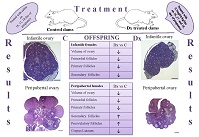
Overexposure to glucocorticoids during fetal development alters fetal organ growth and maturation patterns, which can result in adverse programming outcomes in adulthood. The aim of this study was to determine whether exposure to dexamethasone (Dx) during the fetal period programmed ovary development and function in infant (16-day-old) and peripubertal (38-day-old) female offspring. Pregnant Wistar rats were separated into control and Dx-treated (0.5 mg kg–1) groups and were injected with Dx or an equivalent volume of vehicle on Days 16, 17 and 18 of gestation. Ovaries from 16- and 38-day-old female offspring were prepared for histological and stereological examination. The volume of the ovary and the number of primordial and primary follicles were significantly reduced in prenatally Dx-exposed infant and peripubertal female offspring compared with control offspring. The number of multilaminar follicles was decreased in infant female offspring. In peripubertal females, prenatal exposure to Dx increased the number of multilaminar and large follicles of all classes. Because vaginal opening did not occur up to Day 38 postpartum in the Dx-exposed offspring, the absence of ovulation and corpora lutea is confirmation that the onset of puberty had been delayed. We can conclude that overexposure to glucocorticoids early in life programs ovary development, which may affect fertility in adulthood.
Keywords: development, histology, stereology.
References
Barker, D. J., Hales, C. N., Fall, C. H., Osmond, C., Phipps, K., and Clark, P. M. (1993). Type 2 (non-insulin-dependent) diabetes mellitus, hypertension and hyperlipidaemia (syndrome X): relation to reduced fetal growth. Diabetologia 36, 62–67.| Type 2 (non-insulin-dependent) diabetes mellitus, hypertension and hyperlipidaemia (syndrome X): relation to reduced fetal growth.Crossref | GoogleScholarGoogle Scholar | 8436255PubMed |
Carbone, D. L., Zuloaga, D. G., Hiroi, R., Foradori, C. D., Legare, M. E., and Handa, R. J. (2012). Prenatal dexamethasone exposure potentiates diet-induced hepatosteatosis and decreases plasma IGF-I in a sex-specific fashion. Endocrinology 153, 295–306.
| Prenatal dexamethasone exposure potentiates diet-induced hepatosteatosis and decreases plasma IGF-I in a sex-specific fashion.Crossref | GoogleScholarGoogle Scholar | 22067322PubMed |
Charleston, J. S., Hansen, K. R., Thyer, A. C., Charleston, L. B., Gougeon, A., Siebert, J. R., Soules, M. R., and Klein, N. A. (2007). Estimating human ovarian non-growing follicle number: the application of modern stereology techniques to an old problem. Hum. Reprod. 22, 2103–2110.
| Estimating human ovarian non-growing follicle number: the application of modern stereology techniques to an old problem.Crossref | GoogleScholarGoogle Scholar | 17548367PubMed |
Cottrell, E. C., and Seckl, J. R. (2009). Prenatal stress, glucocorticoids and the programming of adult disease. Front. Behav. Neurosci. 3, 19.
| Prenatal stress, glucocorticoids and the programming of adult disease.Crossref | GoogleScholarGoogle Scholar | 19826624PubMed |
Crowley, P. A. (1995). Antenatal corticosteroid therapy: a meta-analysis of randomized trials, 1972–1994. Am. J. Obstet. Gynecol. 173, 322–335.
| Antenatal corticosteroid therapy: a meta-analysis of randomized trials, 1972–1994.Crossref | GoogleScholarGoogle Scholar | 7631713PubMed |
de Roux, N., Genin, E., Carel, J. C., Matsuda, F., Chaussain, J. L., and Milgrom, E. (2003). Hypogonadotropic hypogonadism due to loss of function of the KiSS1-derived peptide receptor GPR54. Proc. Natl Acad. Sci. USA 100, 10972–10976.
| Hypogonadotropic hypogonadism due to loss of function of the KiSS1-derived peptide receptor GPR54.Crossref | GoogleScholarGoogle Scholar | 12944565PubMed |
de Steenwinkel, F. D. O., Dolhain, R. J. E. M., Hazes, J. M. W., and Hokken-Koelega, A. C. S. (2017). Does prednisone use or disease activity in pregnant women with rheumatoid arthritis influence the body composition of their offspring? Reprod. Toxicol. 71, 118–123.
| Does prednisone use or disease activity in pregnant women with rheumatoid arthritis influence the body composition of their offspring?Crossref | GoogleScholarGoogle Scholar |
Dupont, C., Cordier, A. G., Junien, C., Mandon-Pépin, B., Levy, R., and Chavatte-Palmer, P. (2012). Maternal environment and the reproductive function of the offspring. Theriogenology 78, 1405–1414.
| Maternal environment and the reproductive function of the offspring.Crossref | GoogleScholarGoogle Scholar | 22925651PubMed |
Edson, M. A., Nagaraja, A. K., and Matzuk, M. M. (2009). The mammalian ovary from genesis to revelation. Endocr. Rev. 30, 624–712.
| The mammalian ovary from genesis to revelation.Crossref | GoogleScholarGoogle Scholar | 19776209PubMed |
Findlay, J. K., Hutt, K. J., Hickey, M., and Anderson, R. A. (2015). How is the number of primordial follicles in the ovarian reserve established? Biol. Reprod. 93, 111.
| How is the number of primordial follicles in the ovarian reserve established?Crossref | GoogleScholarGoogle Scholar | 26423124PubMed |
Gaytán, F., Morales, C., Bellido, C., Aguilar, E., and Sánchez-Criado, J. E. (1998). Ovarian follicle macrophages: is follicular atresia in the immature rat a macrophage-mediated event? Biol. Reprod. 58, 52–59.
| Ovarian follicle macrophages: is follicular atresia in the immature rat a macrophage-mediated event?Crossref | GoogleScholarGoogle Scholar | 9472922PubMed |
Gregersen, T. L., and Ulrik, C. S. (2013). Safety of bronchodilators and corticosteroids for asthma during pregnancy: what we know and what we need to do better. J. Asthma Allergy 6, 117–125.
| 24259987PubMed |
Gundersen, H. J., and Jensen, E. B. (1987). The efficiency of systematic sampling in stereology and its prediction. J. Microsc. 147, 229–263.
| The efficiency of systematic sampling in stereology and its prediction.Crossref | GoogleScholarGoogle Scholar | 3430576PubMed |
Irwig, M. S., Fraley, G. S., Smith, J. T., Acohido, B. V., Popa, S. M., Cunningham, M. J., Gottsch, M. L., Clifton, D. K., and Steiner, R. A. (2004). Kisspeptin activation of gonadotropin releasing hormone neurons and regulation of KiSS-1 mRNA in the male rat. Neuroendocrinology 80, 264–272.
| Kisspeptin activation of gonadotropin releasing hormone neurons and regulation of KiSS-1 mRNA in the male rat.Crossref | GoogleScholarGoogle Scholar | 15665556PubMed |
Iwasa, T., Matsuzaki, T., Murakami, M., Fujisawa, S., Kinouchi, R., Gereltsetseg, G., Kuwahara, A., Yasui, T., and Irahara, M. (2010). Effects of intrauterine undernutrition on hypothalamic Kiss1 expression and the timing of puberty in female rats. J. Physiol. 588, 821–829.
| Effects of intrauterine undernutrition on hypothalamic Kiss1 expression and the timing of puberty in female rats.Crossref | GoogleScholarGoogle Scholar | 20083512PubMed |
Iwasa, T., Matsuzaki, T., Murakami, M., Kinouchi, R., Gereltsetseg, G., Yamamoto, S., Kuwahara, A., Yasui, T., and Irahara, M. (2011). Delayed puberty in prenatally glucocorticoid administered female rats occurs independently of the hypothalamic Kiss1–Kiss1r–GnRH system. Int. J. Dev. Neurosci. 29, 183–188.
| Delayed puberty in prenatally glucocorticoid administered female rats occurs independently of the hypothalamic Kiss1–Kiss1r–GnRH system.Crossref | GoogleScholarGoogle Scholar | 21074602PubMed |
Iwasa, T., Matsuzaki, T., Munkhzaya, M., Tungalagsuvd, A., Kawami, T., Murakami, M., Yamasaki, M., Kato, T., Kuwahara, A., Yasui, T., and Irahara, M. (2014). Prenatal exposure to glucocorticoids affects body weight, serum leptin levels, and hypothalamic neuropeptide-Y expression in pre-pubertal female rat offspring. Int. J. Dev. Neurosci. 36, 1–4.
| Prenatal exposure to glucocorticoids affects body weight, serum leptin levels, and hypothalamic neuropeptide-Y expression in pre-pubertal female rat offspring.Crossref | GoogleScholarGoogle Scholar | 24721038PubMed |
Jennes, L. (1989). Prenatal development of the gonadotropin-releasing hormone-containing systems in rat brain. Brain Res. 482, 97–108.
| Prenatal development of the gonadotropin-releasing hormone-containing systems in rat brain.Crossref | GoogleScholarGoogle Scholar | 2650804PubMed |
Kotani, M., Detheux, M., Vandenbogaerde, A., Communi, D., Vanderwinden, J. M., Le Poul, E., Brézillon, S., Tyldesley, R., Suarez-Huerta, N., Vandeput, F., Blanpain, C., Schiffmann, S. N., Vassart, G., and Parmentier, M. (2001). The metastasis suppressor gene KiSS-1 encodes kiss peptins, the natural ligands of the orphan G protein-coupled receptor GPR54. J. Biol. Chem. 276, 34631–34636.
| The metastasis suppressor gene KiSS-1 encodes kiss peptins, the natural ligands of the orphan G protein-coupled receptor GPR54.Crossref | GoogleScholarGoogle Scholar | 11457843PubMed |
Lim, W. L., Soga, T., and Parhar, I. S. (2014). Maternal dexamethasone exposure during pregnancy in rats disrupts gonadotropin-releasing hormone neuronal development in the offspring. Cell Tissue Res. 355, 409–423.
| Maternal dexamethasone exposure during pregnancy in rats disrupts gonadotropin-releasing hormone neuronal development in the offspring.Crossref | GoogleScholarGoogle Scholar | 24374911PubMed |
Manojlović-Stojanoski, M., Ristić, N., Singh, S., and Milosević, V. (2014). Antenatal treatment with glucocorticoids and the HPA axis. J. Med. Biochem. 33, 307–316.
| Antenatal treatment with glucocorticoids and the HPA axis.Crossref | GoogleScholarGoogle Scholar |
Matsui, H., Takatsu, Y., Kumano, S., Matsumoto, H., and Ohtaki, T. (2004). Peripheral administration of metastin induces marked gonadotropin release and ovulation in the rat. Biochem. Biophys. Res. Commun. 320, 383–388.
| Peripheral administration of metastin induces marked gonadotropin release and ovulation in the rat.Crossref | GoogleScholarGoogle Scholar | 15219839PubMed |
Medigović, I., Ristić, N., Trifunović, S., Manojlović-Stojanoski, M., Milošević, V., Zikić, D., and Nestorović, N. (2012). Genistein affects ovarian folliculogenesis: a stereological study. Microsc. Res. Tech. 75, 1691–1699.
| Genistein affects ovarian folliculogenesis: a stereological study.Crossref | GoogleScholarGoogle Scholar | 22927040PubMed |
Miller, P. B., Charleston, J. S., Battaglia, D. E., Klein, N. A., and Soules, M. R. (1997). An accurate, simple method for unbiased determination of primordial follicle number in the primate ovary. Biol. Reprod. 56, 909–915.
| An accurate, simple method for unbiased determination of primordial follicle number in the primate ovary.Crossref | GoogleScholarGoogle Scholar | 9096872PubMed |
Miller, P. B., Charleston, J. S., Battaglia, D. E., Klein, N. A., and Soules, M. R. (1999). Morphometric analysis of primordial follicle number in pigtailed monkey ovaries: symmetry and relationship with age. Biol. Reprod. 61, 553–556.
| Morphometric analysis of primordial follicle number in pigtailed monkey ovaries: symmetry and relationship with age.Crossref | GoogleScholarGoogle Scholar | 10411540PubMed |
Myers, M., Britt, K. L., Wreford, N. G. M., Ebling, F. J. P., and Kerr, J. B. (2004). Methods for quantifying follicular numbers within the mouse ovary. Reproduction 127, 569–580.
| Methods for quantifying follicular numbers within the mouse ovary.Crossref | GoogleScholarGoogle Scholar | 15129012PubMed |
Nathanielsz, P. W. (2006). Animal models that elucidate basic principles of the developmental origins of adult diseases. ILAR J. 47, 73–82.
| Animal models that elucidate basic principles of the developmental origins of adult diseases.Crossref | GoogleScholarGoogle Scholar | 16391433PubMed |
National Research Council (1996). ‘Guide for the Care and Use of Laboratory Animals.’ (The National Academies Press: Washington, DC.)
Navarro, V. M., Castellano, J. M., Fernández-Fernández, R., Barreiro, M. L., Roa, J., Sanchez-Criado, J. E., Aguilar, E., Dieguez, C., Pinilla, L., and Tena-Sempere, M. (2004). Developmental and hormonally regulated messenger ribonucleic acid expression of KiSS-1 and its putative receptor, GPR54, in rat hypothalamus and potent luteinizing hormone-releasing activity of KiSS-1 peptide. Endocrinology 145, 4565–4574.
| Developmental and hormonally regulated messenger ribonucleic acid expression of KiSS-1 and its putative receptor, GPR54, in rat hypothalamus and potent luteinizing hormone-releasing activity of KiSS-1 peptide.Crossref | GoogleScholarGoogle Scholar | 15242985PubMed |
O’Regan, D., Kenyon, C. J., Seckl, J. R., and Holmes, M. C. (2008). Prenatal dexamethasone ‘programmes’ hypotension, but stress-induced hypertension in adult offspring. J. Endocrinol. 196, 343–352.
| Prenatal dexamethasone ‘programmes’ hypotension, but stress-induced hypertension in adult offspring.Crossref | GoogleScholarGoogle Scholar | 18252958PubMed |
Odell, W. D. (1992). Puberty. In ‘Textbook of Endocrinology’. (Eds J. D. Wilson and D. W. Foster.) pp. 1860–1872. (Saunders Co.: Philadelphia.)
Ohtaki, T., Shintani, Y., Honda, S., Matsumoto, H., Hori, A., Kanehashi, K., Terao, Y., Kumano, S., Takatsu, Y., Masuda, Y., Ishibashi, Y., Watanabe, T., Asada, M., Yamada, T., Suenaga, M., Kitada, C., Usuki, S., Kurokawa, T., Onda, H., Nishimura, O., and Fujino, M. (2001). Metastasis suppressor gene KiSS-1 encodes peptide ligand of a G-protein-coupled receptor. Nature 411, 613–617.
| Metastasis suppressor gene KiSS-1 encodes peptide ligand of a G-protein-coupled receptor.Crossref | GoogleScholarGoogle Scholar | 11385580PubMed |
Ortiz, L. A., Quan, A., Weinberg, A., and Baum, M. (2001). Effect of prenatal dexamethasone on rat renal development. Kidney Int. 59, 1663–1669.
| Effect of prenatal dexamethasone on rat renal development.Crossref | GoogleScholarGoogle Scholar | 11318936PubMed |
Osman, P. (1985). Rate and course of atresia during follicular development in the adult cyclic rat. J. Reprod. Fertil. 73, 261–270.
| Rate and course of atresia during follicular development in the adult cyclic rat.Crossref | GoogleScholarGoogle Scholar | 4038517PubMed |
Picut, C. A., Dixon, D., Simons, M. L., Stump, D. G., Parker, G. A., and Remick, A. K. (2015). Postnatal ovary development in the rat: morphologic study and correlation of morphology to neuroendocrine parameters. Toxicol. Pathol. 43, 343–353.
| Postnatal ovary development in the rat: morphologic study and correlation of morphology to neuroendocrine parameters.Crossref | GoogleScholarGoogle Scholar | 25107574PubMed |
Politch, J. A., and Herrenkohl, L. R. (1984). Effects of prenatal stress on reproduction in male and female mice. Physiol. Behav. 32, 95–99.
| Effects of prenatal stress on reproduction in male and female mice.Crossref | GoogleScholarGoogle Scholar | 6718542PubMed |
Poulain, M., Frydman, N., Duquenne, C., N’Tumba-Byn, T., Benachi, A., Habert, R., Rouiller-Fabre, V., and Livera, G. (2012). Dexamethasone induces germ cell apoptosis in the human fetal ovary. J. Clin. Endocrinol. Metab. 97, E1890–E1897.
| Dexamethasone induces germ cell apoptosis in the human fetal ovary.Crossref | GoogleScholarGoogle Scholar | 22802086PubMed |
Rabadán-Diehl, C., and Nathanielsz, P. (2013). From mice to men: research models of developmental programming. J. Dev. Orig. Health Dis. 4, 3–9.
| From mice to men: research models of developmental programming.Crossref | GoogleScholarGoogle Scholar | 23525085PubMed |
Ristić, N., Nestorović, N., Manojlović-Stojanoski, M., Filipović, B., Šošić-Jurjević, B., Milosević, V., and Sekulić, M. (2008). Maternal dexamethasone treatment reduces ovarian follicle number in neonatal rat offspring. J. Microsc. 232, 549–557.
| Maternal dexamethasone treatment reduces ovarian follicle number in neonatal rat offspring.Crossref | GoogleScholarGoogle Scholar | 19094039PubMed |
Ristić, N., Nestorović, N., Manojlović-Stojanoski, M., Medigović, I., Trifunović, S., Šošić-Jurjević, B., and Milošević, V. (2014). Exposure to dexamethasone reduces pituitary volume and gonadotropic cell number in rat fetuses. Acta Histochem. 116, 973–980.
| Exposure to dexamethasone reduces pituitary volume and gonadotropic cell number in rat fetuses.Crossref | GoogleScholarGoogle Scholar | 24816519PubMed |
Ristić, N., Severs, W., Nestorović, N., Jarić, I., Manojlović-Stojanoski, M., Trifunović, S., Pendovski, L., and Milosević, V. (2016). Effects of prenatal dexamethasone on the rat pituitary gland and gonadotropic cells in female offspring. Cells Tissues Organs 201, 148–158.
| Effects of prenatal dexamethasone on the rat pituitary gland and gonadotropic cells in female offspring.Crossref | GoogleScholarGoogle Scholar | 26950885PubMed |
Ristić, N., Nestorović, N., Manojlović-Stojanoski, M., Trifunović, S., Ajdžanović, V., Filipović, B., Pendovski, L., and Milošević, V. (2019). Adverse effect of dexamethasone on development of the fetal rat ovary. Fundam. Clin. Pharmacol. 33, 199–207.
| Adverse effect of dexamethasone on development of the fetal rat ovary.Crossref | GoogleScholarGoogle Scholar | 30216532PubMed |
Sarraj, M. A., and Drummond, A. E. (2012). Mammalian foetal ovarian development: consequences for health and disease. Reproduction 143, 151–163.
| Mammalian foetal ovarian development: consequences for health and disease.Crossref | GoogleScholarGoogle Scholar | 22106406PubMed |
Schwanzel-Fukuda, M., and Pfaff, D. W. (1989). Origin of luteinizing hormone-releasing hormone neurons. Nature 338, 161–164.
| Origin of luteinizing hormone-releasing hormone neurons.Crossref | GoogleScholarGoogle Scholar | 2645530PubMed |
Seminara, S. B., Messager, S., Chatzidaki, E. E., Thresher, R. R., Acierno, J. S., Shagoury, J. K., Bo-Abbas, Y., Kuohung, W., Schwinof, K. M., Hendrick, A. G., Zahn, D., Dixon, J., Kaiser, U. B., Slaugenhaupt, S. A., Gusella, J. F., O’Rahilly, S., Carlton, M. B., Crowley, W. F., Aparicio, S. A., and Colledge, W. H. (2003). The GPR54 gene as a regulator of puberty. N. Engl. J. Med. 349, 1614–1627.
| The GPR54 gene as a regulator of puberty.Crossref | GoogleScholarGoogle Scholar | 14573733PubMed |
Shahab, M., Mastronardi, C., Seminara, S. B., Crowley, W. F., Ojeda, S. R., and Plant, T. M. (2005). Increased hypothalamic GPR54 signaling: a potential mechanism for initiation of puberty in primates. Proc. Natl Acad. Sci. USA 102, 2129–2134.
| Increased hypothalamic GPR54 signaling: a potential mechanism for initiation of puberty in primates.Crossref | GoogleScholarGoogle Scholar | 15684075PubMed |
Smith, J. T., and Waddell, B. J. (2000). Increased fetal glucocorticoid exposure delays puberty onset in postnatal life. Endocrinology 141, 2422–2428.
| Increased fetal glucocorticoid exposure delays puberty onset in postnatal life.Crossref | GoogleScholarGoogle Scholar | 10875242PubMed |
Wear, H. M., McPike, M. J., and Watanabe, K. N. (2016). From primordial germ cells to primordial follicles: a review and visual representation of early ovarian development in mice. J. Ovarian Res. 9, 36.
| From primordial germ cells to primordial follicles: a review and visual representation of early ovarian development in mice.Crossref | GoogleScholarGoogle Scholar | 27329176PubMed |
Wierman, M. E., Kiseljak-Vassiliades, K., and Tobet, S. (2011). Gonadotropin-releasing hormone (GnRH) neuron migration: initiation, maintenance and cessation as critical steps to ensure normal reproductive function. Front. Neuroendocrinol. 32, 43–52.
| Gonadotropin-releasing hormone (GnRH) neuron migration: initiation, maintenance and cessation as critical steps to ensure normal reproductive function.Crossref | GoogleScholarGoogle Scholar | 20650288PubMed |
Wreford, N. G. (1995). Theory and practice of stereological techniques applied to the estimation of cell number and nuclear volume in the testis. Microsc. Res. Tech. 32, 423–436.
| Theory and practice of stereological techniques applied to the estimation of cell number and nuclear volume in the testis.Crossref | GoogleScholarGoogle Scholar | 8563041PubMed |
Yahi, D., Ojo, N. A., Arastus, W., Uchendu, C., Adebayo, A., and Mshelia, G. D. (2017). The use of dexamethasone in animals: implication for fertility, pregnancy and extrapolation of the animal data to humans. Nig. Vet. J. 38, 83–103.