

Time-lapse confocal imaging-induced calcium ion discharge from the cumulus–oocyte complex at the time of cattle oocyte activation
Hanna J. McLennan A B C E , Melanie L. Sutton-McDowall A B C , Sabrina Heng A C D , Andrew D. Abell A C D and Jeremy G. Thompson A B C
A Australian Research Council Centre of Excellence for Nanoscale BioPhotonics, Australia.
B Robinson Research Institute, Adelaide Medical School, University of Adelaide, SA 5005, Australia.
C Institute for Photonics and Advanced Sensing, School of Physical Sciences, University of Adelaide, SA 5005, Australia.
D Department of Chemistry, School of Physical Sciences, University of Adelaide, SA 5005, Australia.
E Corresponding author. Email: hanna.mclennan@adelaide.edu.au
Reproduction, Fertility and Development 32(14) 1223-1238 https://doi.org/10.1071/RD20143
Submitted: 2 June 2020 Accepted: 15 September 2020 Published: 8 October 2020
Abstract
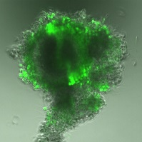
Oocyte activation, the dynamic transformation of an oocyte into an embryo, is largely driven by Ca2+ oscillations that vary in duration and amplitude across species. Previous studies have analysed intraoocyte Ca2+ oscillations in the absence of the oocyte’s supporting cumulus cells. Therefore, it is unknown whether cumulus cells also produce an ionic signal that reflects fertilisation success. Time-lapse confocal microscopy and image analysis on abattoir-derived cattle cumulus–oocyte complexes coincubated with spermatozoa revealed a distinct discharge of fluorescence from the cumulus vestment. This study demonstrated that this Ca2+ fluorescence discharge was an artefact induced by the imaging procedure independently of oocyte activation success. The fluorescence discharge was a direct result of cumulus cell membrane integrity loss, and future studies should consider the long-term effect of fluorescent labels on cells in time-lapse imaging. However, this study also demonstrated that the distinctive pattern of a coordinated fluorescence discharge was associated with both the presence of spermatozoa and subsequent embryo development to the morula stage, which was affected by Ca2+ chelation and a reduction in the active efflux of the fluorophore. This indicates that the cumulus vestment may have a relationship with oocyte activation at and beyond fertilisation that requires further investigation.
Keywords: calcium oscillations, confocal imaging, embryo development, fertilisation, fluorescent probes, oocyte activation.
References
Albertini, D. F., Combelles, C. M., Benecchi, E., and Carabatsos, M. J. (2001). Cellular basis for paracrine regulation of ovarian follicle development. Reproduction 121, 647–653.| Cellular basis for paracrine regulation of ovarian follicle development.Crossref | GoogleScholarGoogle Scholar | 11427152PubMed |
Ben-Yosef, D., and Shalgi, R. (1998). Early ionic events in activation of the mammalian egg. Rev. Reprod. 3, 96–103.
| Early ionic events in activation of the mammalian egg.Crossref | GoogleScholarGoogle Scholar | 9685188PubMed |
Carroll, J., Swann, K., Whittingham, D., and Whitaker, M. (1994). Spatiotemporal dynamics of intracellular [Ca2+]i oscillations during the growth and meiotic maturation of mouse oocytes. Development 120, 3507–3517.
| 7821218PubMed |
Chiba, K., Kado, R. T., and Jaffe, L. A. (1990). Development of calcium release mechanisms during starfish oocyte maturation. Dev. Biol. 140, 300–306.
| Development of calcium release mechanisms during starfish oocyte maturation.Crossref | GoogleScholarGoogle Scholar | 2373255PubMed |
Cuthbertson, K. S. R., Whittingham, D. G., and Cobbold, P. H. (1981). Free Ca2+ increases in exponential phases during mouse oocyte activation. Nature 294, 754–757.
| Free Ca2+ increases in exponential phases during mouse oocyte activation.Crossref | GoogleScholarGoogle Scholar |
Dale, B., Defelice, L. J., and Ehrenstein, G. (1985). Injection of a soluble sperm fraction into sea urchin eggs triggers the cortical reaction. Experientia 41, 1068–1070.
| Injection of a soluble sperm fraction into sea urchin eggs triggers the cortical reaction.Crossref | GoogleScholarGoogle Scholar | 4018233PubMed |
Di Virgilio, F., Steinberg, T. H., and Silverstein, S. C. (1990). Inhibition of Fura-2 sequestration and secretion with organic anion transport blockers. Cell. Calcium. 11, 57–62.
| Inhibition of Fura-2 sequestration and secretion with organic anion transport blockers.Crossref | GoogleScholarGoogle Scholar | 2191781PubMed |
Dumollard, R., Carroll, J., Dupont, G., and Sardet, C. (2002). Calcium wave pacemakers in eggs. J. Cell Sci. 115, 3557–3564.
| Calcium wave pacemakers in eggs.Crossref | GoogleScholarGoogle Scholar | 12186942PubMed |
Dumollard, R., Hammar, K., Porterfield, M., Smith, P. J., Cibert, C., Rouviere, C., and Sardet, C. (2003). Mitochondrial respiration and Ca2+ waves are linked during fertilization and meiosis completion. Development 130, 683–692.
| Mitochondrial respiration and Ca2+ waves are linked during fertilization and meiosis completion.Crossref | GoogleScholarGoogle Scholar | 12505999PubMed |
Duncan, F. E., Que, E. L., Zhang, N., Feinberg, E. C., O’Halloran, T. V., and Woodruff, T. K. (2016). The zinc spark is an inorganic signature of human egg activation. Sci. Rep. 6, 24737.
| The zinc spark is an inorganic signature of human egg activation.Crossref | GoogleScholarGoogle Scholar | 27113677PubMed |
Eppig, J. J. (1991). Intercommunication between mammalian oocytes and companion somatic cells. BioEssays 13, 569–574.
| Intercommunication between mammalian oocytes and companion somatic cells.Crossref | GoogleScholarGoogle Scholar | 1772412PubMed |
Fatehi, A. N., Zeinstra, E. C., Kooij, R. V., Colenbrander, B., and Bevers, M. M. (2002). Effect of cumulus cell removal of in vitro matured bovine oocytes prior to in vitro fertilization on subsequent cleavage rate. Theriogenology 57, 1347–1355.
| Effect of cumulus cell removal of in vitro matured bovine oocytes prior to in vitro fertilization on subsequent cleavage rate.Crossref | GoogleScholarGoogle Scholar | 12013454PubMed |
Fujiwara, T., Nakada, K., Shirakawa, H., and Miyazaki, S. (1993). Development of inositol trisphosphate-induced calcium release mechanism during maturation of hamster oocytes. Dev. Biol. 156, 69–79.
| Development of inositol trisphosphate-induced calcium release mechanism during maturation of hamster oocytes.Crossref | GoogleScholarGoogle Scholar | 8383620PubMed |
Fukuda, Y., Okada, O., and Toyoda, Y. (1972). Studies on the fertilization of mouse eggs in vitro III. Fertilization of denuded eggs by capacitated spermatozoa. Jpn. J. Anim. Reprod. 18, 73–77.
| Studies on the fertilization of mouse eggs in vitro III. Fertilization of denuded eggs by capacitated spermatozoa.Crossref | GoogleScholarGoogle Scholar |
Geshi, M., Takenouchi, N., Yamauchi, N., and Nagai, T. (2000). Effects of sodium pyruvate in nonserum maturation medium on maturation, fertilization, and subsequent development of bovine oocytes with or without cumulus cells. Biol. Reprod. 63, 1730–1734.
| Effects of sodium pyruvate in nonserum maturation medium on maturation, fertilization, and subsequent development of bovine oocytes with or without cumulus cells.Crossref | GoogleScholarGoogle Scholar | 11090443PubMed |
Gilula, N. B., Epstein, M. L., and Beers, W. H. (1978). Cell-to-cell communication and ovulation: a study of the cumulus–oocyte complex. J. Cell Biol. 78, 58–75.
| Cell-to-cell communication and ovulation: a study of the cumulus–oocyte complex.Crossref | GoogleScholarGoogle Scholar | 670298PubMed |
Grynkiewicz, G., Poenie, M., and Tsien, R. Y. (1985). A new generation of Ca2+ indicators with greatly improved fluorescence properties. J. Biol. Chem. 260, 3440–3450.
| 3838314PubMed |
He, C. L., Damiani, P., Parys, J. B., and Fissore, R. A. (1997). Calcium, calcium release receptors, and meiotic resumption in bovine oocytes. Biol. Reprod. 57, 1245–1255.
| Calcium, calcium release receptors, and meiotic resumption in bovine oocytes.Crossref | GoogleScholarGoogle Scholar | 9369194PubMed |
Homa, S. T., Carroll, J., and Swann, K. (1993). The role of calcium in mammalian oocyte maturation and egg activation. Hum. Reprod. 8, 1274–1281.
| 8408526PubMed |
Hyttel, P. (1987). Bovine cumulus–oocyte disconnection in vitro. Anat. Embryol. (Berl.) 176, 41–44.
| Bovine cumulus–oocyte disconnection in vitro.Crossref | GoogleScholarGoogle Scholar | 3605648PubMed |
Jones, K. T. (2005). Mammalian egg activation: from Ca2+ spiking to cell cycle progression. Reproduction 130, 813–823.
| Mammalian egg activation: from Ca2+ spiking to cell cycle progression.Crossref | GoogleScholarGoogle Scholar | 16322541PubMed |
Kim, A. M., Bernhardt, M. L., Kong, B. Y., Ahn, R. W., Vogt, S., Woodruff, T. K., and O’Halloran, T. V. (2011). Zinc sparks are triggered by fertilization and facilitate cell cycle resumption in mammalian eggs. ACS Chem. Biol. 6, 716–723.
| Zinc sparks are triggered by fertilization and facilitate cell cycle resumption in mammalian eggs.Crossref | GoogleScholarGoogle Scholar | 21526836PubMed |
Kline, D., and Kline, J. T. (1992). Repetitive calcium transients and the role of calcium in exocytosis and cell cycle activation in the mouse egg. Dev. Biol. 149, 80–89.
| Repetitive calcium transients and the role of calcium in exocytosis and cell cycle activation in the mouse egg.Crossref | GoogleScholarGoogle Scholar | 1728596PubMed |
Kline, D., Mehlmann, L., Fox, C., and Terasaki, M. (1999). The cortical endoplasmic reticulum (ER) of the mouse egg: localization of ER clusters in relation to the generation of repetitive calcium waves. Dev. Biol. 215, 431–442.
| The cortical endoplasmic reticulum (ER) of the mouse egg: localization of ER clusters in relation to the generation of repetitive calcium waves.Crossref | GoogleScholarGoogle Scholar | 10545249PubMed |
Liu, M. (2011). The biology and dynamics of mammalian cortical granules. Reprod. Biol. Endocrinol. 9, 149.
| The biology and dynamics of mammalian cortical granules.Crossref | GoogleScholarGoogle Scholar | 22088197PubMed |
Lorton, S. P., and First, N. L. (1979). Hyaluronidase does not disperse the cumulus oophorus surrounding bovine ova. Biol. Reprod. 21, 301–308.
| Hyaluronidase does not disperse the cumulus oophorus surrounding bovine ova.Crossref | GoogleScholarGoogle Scholar | 486657PubMed |
Macaulay, A. D., Gilbert, I., Scantland, S., Fournier, E., Ashkar, F., Bastien, A., Saadi, H. A. S., Gagne, D., Sirard, M. A., Khandjian, E. W., Richard, F. J., Hyttel, P., and Robert, C. (2016). Cumulus cell transcripts transit to the bovine oocyte in preparation for maturation. Biol. Reprod. 94, 11.
| Cumulus cell transcripts transit to the bovine oocyte in preparation for maturation.Crossref | GoogleScholarGoogle Scholar |
McLennan, H. J., Saini, A., Sylvia, G. M., Schartner, E. P., Dunning, K. R., Purdey, M. S., Monro, T. M., Abell, A. D., and Thompson, J. G. (2019). A biophotonic approach to measure pH in small volumes in vitro: quantifiable differences in metabolic flux around the cumulus–oocyte-complex (COC). J. Biophotonics 13, e201960038.
| A biophotonic approach to measure pH in small volumes in vitro: quantifiable differences in metabolic flux around the cumulus–oocyte-complex (COC).Crossref | GoogleScholarGoogle Scholar | 31725948PubMed |
Mehlmann, L. M., and Kline, D. (1994). Regulation of intracellular calcium in the mouse egg: calcium release in response to sperm or inositol trisphosphate is enhanced after meiotic maturation. Biol. Reprod. 51, 1088–1098.
| Regulation of intracellular calcium in the mouse egg: calcium release in response to sperm or inositol trisphosphate is enhanced after meiotic maturation.Crossref | GoogleScholarGoogle Scholar | 7888488PubMed |
Miyazaki, S., and Igusa, Y. (1981). Fertilization potential in golden hamster eggs consists of recurring hyperpolarizations. Nature 290, 702–704.
| Fertilization potential in golden hamster eggs consists of recurring hyperpolarizations.Crossref | GoogleScholarGoogle Scholar | 6894326PubMed |
Mizuno, S., Ishikawa, Y., Matsumoto, H., Sato, M., Ida, M., Fukuda, A., and Morimoto, Y. (2019). The timing of cumulus cell removal for intracytoplasmic sperm injection influences the capability of embryonic development. Reprod. Med. Biol. 18, 111–117.
| The timing of cumulus cell removal for intracytoplasmic sperm injection influences the capability of embryonic development.Crossref | GoogleScholarGoogle Scholar | 30655729PubMed |
Morado, S., Cetica, P., Beconi, M., Thompson, J. G., and Dalvit, G. (2013). Reactive oxygen species production and redox state in parthenogenetic and sperm-mediated bovine oocyte activation. Reproduction 145, 471–478.
| Reactive oxygen species production and redox state in parthenogenetic and sperm-mediated bovine oocyte activation.Crossref | GoogleScholarGoogle Scholar | 23630331PubMed |
Parrington, J., Arnoult, C., and Fissore, R. A. (2018). The eggstraordinary story of how life begins. Mol. Reprod. Dev. 86, 4–19.
| The eggstraordinary story of how life begins.Crossref | GoogleScholarGoogle Scholar | 30411426PubMed |
Potter, S. M. (1996). Vital imaging: two photons are better than one. Curr. Biol. 6, 1595–1598.
| Vital imaging: two photons are better than one.Crossref | GoogleScholarGoogle Scholar | 8994823PubMed |
Que, E. L., Bleher, R., Duncan, F. E., Kong, B. Y., Gleber, S. C., Vogt, S., Chen, S., Garwin, S. A., Bayer, A. R., Dravid, V. P., Woodruff, T. K., and O’Halloran, T. V. (2015). Quantitative mapping of zinc fluxes in the mammalian egg reveals the origin of fertilization-induced zinc sparks. Nat. Chem. 7, 130–139.
| Quantitative mapping of zinc fluxes in the mammalian egg reveals the origin of fertilization-induced zinc sparks.Crossref | GoogleScholarGoogle Scholar | 25615666PubMed |
Que, E. L., Duncan, F. E., Bayer, A. R., Philips, S. J., Roth, E. W., Bleher, R., Gleber, S. C., Vogt, S., Woodruff, T. K., and O’Halloran, T. V. (2017). Zinc sparks induce physiochemical changes in the egg zona pellucida that prevent polyspermy. Integr. Biol. 9, 135–144.
| Zinc sparks induce physiochemical changes in the egg zona pellucida that prevent polyspermy.Crossref | GoogleScholarGoogle Scholar |
Que, E. L., Duncan, F. E., Lee, H. C., Hornick, J. E., Vogt, S., Fissore, R. A., O’Halloran, T. V., and Woodruff, T. K. (2019). Bovine eggs release zinc in response to parthenogenetic and sperm-induced egg activation. Theriogenology 127, 41–48.
| Bovine eggs release zinc in response to parthenogenetic and sperm-induced egg activation.Crossref | GoogleScholarGoogle Scholar | 30639695PubMed |
Robertson, S. A. (2005). Seminal plasma and male factor signalling in the female reproductive tract. Cell Tissue Res. 322, 43–52.
| Seminal plasma and male factor signalling in the female reproductive tract.Crossref | GoogleScholarGoogle Scholar | 15909166PubMed |
Russell, D. L., Gilchrist, R. B., Brown, H. M., and Thompson, J. G. (2016). Bidirectional communication between cumulus cells and the oocyte: old hands and new players? Theriogenology 86, 62–68.
| Bidirectional communication between cumulus cells and the oocyte: old hands and new players?Crossref | GoogleScholarGoogle Scholar | 27160446PubMed |
Schjenken, J. E., and Robertson, S. A. (2015). Seminal fluid signalling in the female reproductive tract: implications for reproductive success and offspring health. In ‘Male Role in Pregnancy Loss and Embryo Implantation Failure’. Vol. 868. (Ed. R. Bronson.) pp. 127–158. (Springer-Verlag: Berlin.)
Schultz, R. M., and Kopf, G. S. (1995). Molecular basis of mammalian egg activation. Curr. Top. Dev. Biol. 30, 21–62.
| Molecular basis of mammalian egg activation.Crossref | GoogleScholarGoogle Scholar | 7555047PubMed |
Silvestre, F., Fissore, R. A., Tosti, E., and Boni, R. (2012). Ca2+ rise at in vitro maturation in bovine cumulus–oocyte complexes. Mol. Reprod. Dev. 79, 369–379.
| Ca2+ rise at in vitro maturation in bovine cumulus–oocyte complexes.Crossref | GoogleScholarGoogle Scholar | 22431451PubMed |
Sokka, A. L., Putkonen, N., Mudo, G., Pryazhnikov, E., Reijonen, S., Khiroug, L., Belluardo, N., Lindholm, D., and Korhonen, L. (2007). Endoplasmic reticulum stress inhibition protects against excitotoxic neuronal injury in the rat brain. J. Neurosci. 27, 901–908.
| Endoplasmic reticulum stress inhibition protects against excitotoxic neuronal injury in the rat brain.Crossref | GoogleScholarGoogle Scholar | 17251432PubMed |
Stricker, S. A. (1999). Comparative biology of calcium signaling during fertilization and egg activation in animals. Dev. Biol. 211, 157–176.
| Comparative biology of calcium signaling during fertilization and egg activation in animals.Crossref | GoogleScholarGoogle Scholar | 10395780PubMed |
Sun, F. Z., Bradshaw, J. P., Galli, C., and Moor, R. M. (1994). Changes in intracellular calcium concentration in bovine oocytes following penetration by spermatozoa. J. Reprod. Fertil. 101, 713–719.
| Changes in intracellular calcium concentration in bovine oocytes following penetration by spermatozoa.Crossref | GoogleScholarGoogle Scholar | 7966030PubMed |
Sutton, M. L., Gilchrist, R. B., and Thompson, J. G. (2003). Effects of in-vivo and in-vitro environments on the metabolism of the cumulus–oocyte complex and its influence on oocyte developmental capacity. Hum. Reprod. Update 9, 35–48.
| Effects of in-vivo and in-vitro environments on the metabolism of the cumulus–oocyte complex and its influence on oocyte developmental capacity.Crossref | GoogleScholarGoogle Scholar | 12638780PubMed |
Sutton-McDowall, M. L., Gilchrist, R. B., and Thompson, J. G. (2004). Cumulus expansion and glucose utilisation by bovine cumulus–oocyte complexes during in vitro maturation: the influence of glucosamine and follicle-stimulating hormone. Reproduction 128, 313–319.
| Cumulus expansion and glucose utilisation by bovine cumulus–oocyte complexes during in vitro maturation: the influence of glucosamine and follicle-stimulating hormone.Crossref | GoogleScholarGoogle Scholar | 15333782PubMed |
Sutton-McDowall, M. L., Gilchrist, R. B., and Thompson, J. G. (2010). The pivotal role of glucose metabolism in determining oocyte developmental competence. Reproduction 139, 685–695.
| The pivotal role of glucose metabolism in determining oocyte developmental competence.Crossref | GoogleScholarGoogle Scholar | 20089664PubMed |
Swann, K. (1990). A cytosolic sperm factor stimulates repetitive calcium increases and mimics fertilization in hamster eggs. Development 110, 1295–1302.
| 2100264PubMed |
Swann, K., and Whitaker, M. (1986). The part played by inositol trisphosphate and calcium in the propagation of the fertilization wave in sea-urchin eggs. J. Cell Biol. 103, 2333–2342.
| The part played by inositol trisphosphate and calcium in the propagation of the fertilization wave in sea-urchin eggs.Crossref | GoogleScholarGoogle Scholar | 3491080PubMed |
Takahashi, K., Inanami, O., and Kuwabara, M. (1999). Effects of intracellular calcium chelator BAPTA-AM on radiation-induced apoptosis regulated by activation of SAPK/JNK and caspase-3 in MOLT-4 cells. Int. J. Radiat. Biol. 75, 1099–1105.
| Effects of intracellular calcium chelator BAPTA-AM on radiation-induced apoptosis regulated by activation of SAPK/JNK and caspase-3 in MOLT-4 cells.Crossref | GoogleScholarGoogle Scholar | 10528917PubMed |
Thévenaz, P., Ruttimann, U. E., and Unser, M. (1998). A pyramid approach to subpixel registration based on intensity. IEEE Trans. Image Process. 7, 27–41.
| A pyramid approach to subpixel registration based on intensity.Crossref | GoogleScholarGoogle Scholar | 18267377PubMed |
Thompson, J. G. E., and Wales, R. G. (1987). Observations on the loss of the cellular vestment surrounding superovulated sheep oocytes. Anim. Reprod. Sci. 15, 169–175.
| Observations on the loss of the cellular vestment surrounding superovulated sheep oocytes.Crossref | GoogleScholarGoogle Scholar |
Turner, K., and Horobin, R. W. (1997). Permeability of the mouse zona pellucida: a structure-staining-correlation model using coloured probes. J. Reprod. Fertil. 111, 259–265.
| Permeability of the mouse zona pellucida: a structure-staining-correlation model using coloured probes.Crossref | GoogleScholarGoogle Scholar | 9462294PubMed |
Tuszyński, J. A., Portet, S., Dixon, J. M., Luxford, C., and Cantiello, H. F. (2004). Ionic wave propagation along actin filaments. Biophys. J. 86, 1890–1903.
| Ionic wave propagation along actin filaments.Crossref | GoogleScholarGoogle Scholar | 15041636PubMed |
Van Soom, A., Tanghe, S., De Pauw, I., Maes, D., and de Kruif, A. (2002). Function of the cumulus oophorus before and during mammalian fertilization. Reprod. Domest. Anim. 37, 144–151.
| Function of the cumulus oophorus before and during mammalian fertilization.Crossref | GoogleScholarGoogle Scholar | 12071888PubMed |
Vergara, G. J., Irwin, M. H., Moffatt, R. J., and Pinkert, C. A. (1997). In vitro fertilization in mice: strain differences in response to superovulation protocols and effect of cumulus cell removal. Theriogenology 47, 1245–1252.
| In vitro fertilization in mice: strain differences in response to superovulation protocols and effect of cumulus cell removal.Crossref | GoogleScholarGoogle Scholar | 16728073PubMed |
Webb, R. J., Bains, H., Cruttwell, C., and Carroll, J. (2002). Gap-junctional communication in mouse cumulus-oocyte complexes: implications for the mechanism of meiotic maturation. Reproduction 123, 41–52.
| Gap-junctional communication in mouse cumulus-oocyte complexes: implications for the mechanism of meiotic maturation.Crossref | GoogleScholarGoogle Scholar | 11869185PubMed |
Yeste, M., Jones, C., Amdani, S. N., Patel, S., and Coward, K. (2016). Oocyte activation deficiency: a role for an oocyte contribution? Hum. Reprod. Update 22, 23–47.
| Oocyte activation deficiency: a role for an oocyte contribution?Crossref | GoogleScholarGoogle Scholar | 26346057PubMed |
Yoon, S. B., Choi, S. A., Sim, B. W., Kim, J. S., Mun, S. E., Jeong, P. S., Yang, H. J., Lee, Y., Park, Y. H., Song, B. S., Kim, Y. H., Jeong, K. J., Huh, J. W., Lee, S. R., Kim, S. U., and Chang, K. T. (2014). Developmental competence of bovine early embryos depends on the coupled response between oxidative and endoplasmic reticulum stress. Biol. Reprod. 90, 10.
| Developmental competence of bovine early embryos depends on the coupled response between oxidative and endoplasmic reticulum stress.Crossref | GoogleScholarGoogle Scholar |
Zhang, N., Duncan, F. E., Que, E. L., O’Halloran, T. V., and Woodruff, T. K. (2016). The fertilization-induced zinc spark is a novel biomarker of mouse embryo quality and early development. Sci. Rep. 6, 22772.
| The fertilization-induced zinc spark is a novel biomarker of mouse embryo quality and early development.Crossref | GoogleScholarGoogle Scholar | 26987302PubMed |