29 COMPARISON OF TERM PLACENTAS IN CLONED AND CONTROL PREGNANCIES IN CATTLE
C.A. Batchelder A , M. Bertolini A , K.A. Hoffert A , J.B. Mason A , A.L. Moyer A , S.G. Petkov A and G.B. Anderson AADepartment of Animal Science, University of California-Davis, Davis, CA 95616, USA. Email: cabatchelder@ucdavis.edu
Reproduction, Fertility and Development 17(2) 164-165 https://doi.org/10.1071/RDv17n2Ab29
Submitted: 1 August 2004 Accepted: 1 October 2004 Published: 1 January 2005
Abstract
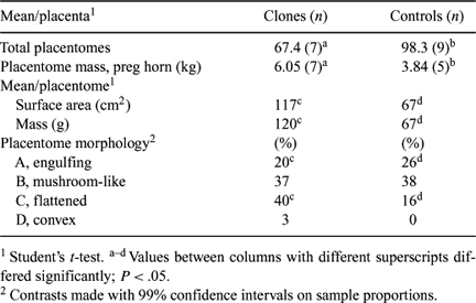
Somatic cell nuclear transfer is associated with high incidence of fetal loss, late-term pregnancy complications, perinatal mortality, and abnormal placental development. Several groups have described abnormalities of early and mid-gestation cloned placentas (Hill et al. 2000 Biol. Reprod. 63, 1787–1794; Lee et al. 2004 Biol. Reprod. 70, 1–11). The objective of our study was to characterize differences in the placentas of clones and control calves at term delivery. Clones were produced from ovarian cell lines from two donors (Holstein, n = 5; Hereford, n = 2). Breed-matched controls included AI (Holstein, n = 3) and embryo transfer (Holstein, n = 3; Hereford n = 3) calves. All calves were delivered alive with no visible birth defects between Days 273 and 280 of gestation, and placentas were recovered for measurement and morphological analysis. When possible, pregnancies were delivered via caesarian section, and the entire uterus was recovered for classification of anatomical shape of placentomes. Each placentome was measured, weighed, and classified by type as (A) engulfing mushroom-like; (B) sub-engulfing mushroom-like; (C) flattened, non-engulfing; and (D) convex (adapted from Penninga and Longo 1998 Placenta 19, 187–193, for sheep). Mean number of placentomes per placenta was significantly greater in controls than clones, while total mass of placentomes in the pregnant horn was significantly greater in clones than in controls (Table 1). Total surface area of placentomes in the pregnant horn tended to be larger and more variable in clones (range: 2710–7450 cm2) than in controls (range: 3120–5030 cm2; P < 0.10). A two-fold increase was observed in cloned placentas, as compared with control placentas, in mean surface area per placentome and mass per placentome. Anatomically, cloned placentas differed from controls in the percentage of placentomes classified Type A (controls > clones) and Type C (clones > controls). Other abnormalities noted in cloned placentas included moderate to severe edema, teratomas, enlarged vessels, and large areas devoid of placentation. All clones and 2/9 controls displayed enlarged umbilical vessels. Significant placental abnormalities were observed in all cloned pregnancies.

|


