293 QUANTITATIVE TESTICULAR HISTOLOGY IN THE COMMON WOMBAT (VOMBATUS URSINUS)
C.A. MacCallum B and S.D. Johnston AA School of Veterinary Science, Queensland University, Queensland, Australia;;
B Western Plains Zoo, New South Wales, Australia. email: cmaccallum@zoo.nsw.gov.au
Reproduction, Fertility and Development 16(2) 266-266 https://doi.org/10.1071/RDv16n1Ab293
Submitted: 1 August 2003 Accepted: 1 October 2003 Published: 2 January 2004
Abstract
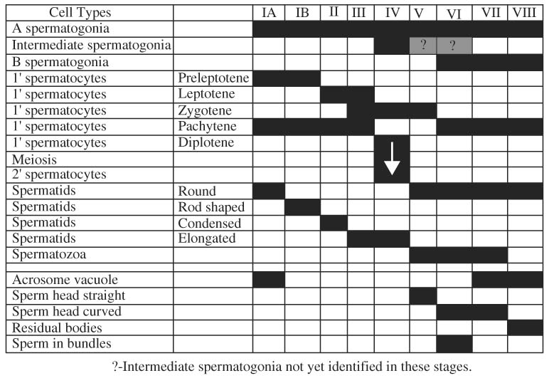
The seminiferous epithelium cycle of the male Common Wombat ( Vombatus ursinus) is documented here for the first time. Testicular material was obtained from 10 common wombats from the Southern Highlands of NSW in June (n = 5) and November (n = 5), fixed in Bouins solution and prepared for standard histological processing. Eight stages of the seminiferous cycle where identified based upon relative cellular associations and development of the spermatid during spermiogenesis. Stage 1 was further subdivided into 1A and 1B based on changes in shape of the spermatid nucleus. The relative frequency of each stage was also calculated using observations from 500 seminiferous tubule cross-sections as was the proportion of the various testicular tissue types. The relative proportions of the various stages of the seminiferous epithelial cycle in the common wombat testis were: Stage 1A, 4.0 ± 0.5; Stage 1B, 4.2 ± 0.4; Stage 2, 21.3 ± 1.9; Stage 3, 15.4 ± 1.2; Stage 4, 16.8 ± 1.2; Stage 5, 11.1 ± 1.5; Stage 6, 13.7 ± 1.5; Stage 7, 7.3 ± 0.6; Stage 8, 6.1 ± 0.6. Relative proportions of the various tissue types observed in testis included: seminiferous tubules (41.5% ± 4.1); seminiferous tubule lumen (33.3 ± 3.4%); leydig cells (14.6 ± 1.1%); connective tissue (10.4 ± 0.9%) and blood vessels (0.2 ± 0.03%).

|


