48 PREGNANCY AND FETAL CHARACTERISTICS AFTER TRANSFER OF VITRIFIED IN VIVO AND CLONED BOVINE EMBRYOS
P. Lonergan A , A.C.O. Evans A , E. Boland A , D. Rizos A , L.-Y. Sung B , F. Du B C , S. Chaubal B , S. Fair A , E. Scraggs A , P. Duffy A , J. Xu A , X. Yang B and X.C. Tian BA Department of Animal Science and Centre for Integrative Biology, Conway Institute for Biomolecular and Biomedical Research, University College Dublin, Newcastle, Co. Dublin, Ireland
B Department of Animal Science, Center for Regenerative Biology, University of Connecticut, Storrs, CT, USA
C Evergen Biotechnologies, Inc., Storrs, CT, USA. Email: pat.lonergan@ucd.ie
Reproduction, Fertility and Development 17(2) 173-174 https://doi.org/10.1071/RDv17n2Ab48
Submitted: 1 August 2004 Accepted: 1 October 2004 Published: 1 January 2005
Abstract
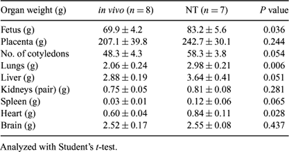
This study was conducted to examine pregnancy progression and fetal characteristics following transfer of vitrified bovine nuclear transfer (NT) vs. in vivo-derived (Vivo) embryos. Nuclear transfer was conducted using skin fibroblast cells derived from cultured ear explants taken from an elite Holstein-Friesian dairy cow. Expanding and hatching blastocysts on Day 7 were vitrified using liquid nitrogen surface vitrification (Shen et al. 2004 Reprod. Fertil. Dev. 16, 158). Day 7 in vivo embryos, produced using standard superovulation procedures applied to Holstein-Friesian heifers (n = 6), were vitrified in the same way. Following warming, embryos were transferred to synchronized recipients (2 per recipient; NT: n = 81 recipients; Vivo: n = 20 recipients). Pregnancies were monitored by ultrasound scanning on Days 25, 45, and 75, and a sample of animals was slaughtered at each time point to recover the fetus/placenta for further analyses. On Day 25, the entire conceptus was weighed together; on Day 45, the fetus and placenta were weighed separately; on Day 75, the fetus was dissected and the major organs were weighed. Significantly more animals remained pregnant after transfer of in vivo-derived embryos than NT embryos at all time points: Day 25 (95.0 vs. 61.7%, P < 0.01), Day 45 (93.3 vs. 39.7%, P < 0.001), and Day 75 (90.9 vs. 19.8%, P < 0.001). There was no significant difference (P = 0.10) in the weight of the conceptus on Day 25 from NT transfers (1.14 ± 0.23 g, n = 8) vs. Vivo transfers (0.75 ± 0.19 g, n = 8). On Day 45, there was no significant difference in the weight of either fetus (P = 0.393) or membranes (P = 0.167) between NT embryos (fetus: 2.76 ± 0.40 g, n = 12; membranes 59.0 ± 10.0 g, n = 11) or in vivo-derived embryos (fetus: 2.60 ± 0.15 g, n = 6; membranes 41.8 ± 5.2 g, n = 4). However, on Day 75, the weights of the fetus and several of the major organs were heavier from NT embryos (Table 1). These data suggest that the large calf syndrome is manifested after Day 45.

|
This work was supported by the U.S. Department of Agriculture.


