97 PRELIMINARY DESCRIPTIVE STUDY OF EQUINE PLACENTA GENERATED AFTER TRANSFER OF IN VIVO- AND IN VITRO-PRODUCED EMBRYOS
A. Lanci A , J. Mariella A , B. Merlo A , C. Castagnetti A and E. Iacono ADepartment of Veterinary Medical Sciences, Ozzano dell’Emilia, Bologna, Italy
Reproduction, Fertility and Development 29(1) 156-156 https://doi.org/10.1071/RDv29n1Ab97
Published: 2 December 2016
Abstract
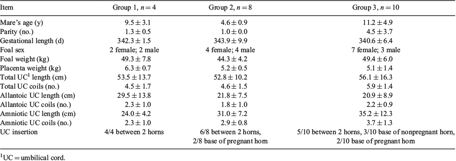
Placental changes associated with artificial reproductive technologies have been described in several species, but little information is available in horses. Joy et al. (2012) reported that human placentas from intracytoplasmic sperm injection derived embryos were heavier and thicker than those produced after natural conception. Despite the most growing interest and efficiency of artificial reproductive technologies in equine species, only recently, Pozor et al. (2016) described placental abnormalities in pregnancies generated by somatic cell NT, but there are no studies on equine placenta generated by intracytoplasmic sperm injection and traditional embryo transfer. In the present preliminary study, macroscopic differences of placentas generated after transfer of in vitro- or in vivo-produced embryos were registered. Twelve Standardbred recipient mares with pregnancy generated after transfer of in vivo-derived (Group 1) and in vitro-derived (Group 2) embryos were enrolled; 10 Standardbred mares with pregnancy derived by traditional AI were included as control (Group 3). All pregnancies were physiological, and newborn foals were healthy. Mare age, parity, length of pregnancy, gross evaluation and weight of placenta, total length of umbilical cord (UC), length of UC, number of UC coils, foal sex, and weight at birth were registered. Collected data are listed in Table 1 and are expressed as mean ± standard deviation. Differences between groups were evaluated by 1-way ANOVA, and the difference in proportion of overweight placentas was evaluated with the Fisher test. The gross evaluation of placenta revealed 8/12 placentas (2/4 Group 1; 6/8 Group 2) were heavier than 11% (Madigan, 1997) due to oedema of the chorioallantois. No overweight placentas were registered in Group 3. In Group 1, 1/4 placentas had villous hypoplasia, and in Group 2, 1/8 placentas had cystic pouches on the UC. There were no significant differences among groups. However, the proportion of overweight placentas between Group 2 (6/8) and Group 3 (0/10) approached significance (P = 0.06). Although preliminary, the results of the present study suggest that production of equine embryos in vitro may lead to alterations in placental development. Several studies in cattle and sheep have suggested that alterations in the placentas of pregnancies derived from in vitro-produced embryos are related to effects of culture on epigenetic regulation. Less is known in the horse about the effects of in vitro embryo production on placental development; thus, further research in this area is necessary.

|


