First record of Cercospora basellae-albae from the Philippines
M. Mahamuda Begum A and C. J. R. Cumagun A B CA Crop Protection Cluster, College of Agriculture, University of the Philippines Los Baños, College, Laguna 4031, Philippines.
B Current address. Laboratory of Plant Pathology, Graduate School of Agricultural Science, Kobe University, Nada, Kobe 657-8501, Japan.
C Corresponding author. Email: cumagun@bear.kobe-u.ac.jp
Australasian Plant Disease Notes 5(1) 115-116 https://doi.org/10.1071/DN10042
Submitted: 10 September 2010 Accepted: 29 September 2010 Published: 11 October 2010
Abstract
Necrotic spots on both young and mature leaves of Basella alba cv. Rubra were found at different sites near Los Baños, Laguna Province in the Philippines. The spots were colonised by Cercospora basellae-albae. This is the first record of Cercospora basellae-albae from the Philippines.
Additional keywords: Cercospora basellae-albae, Basella alba, Basella rubra.
Introduction
Basella alba (Basellaceae), also known as Indian spinach, Ceylon spinach, vine spinach, Malabar spinach or Malabar nightshade is an attractive perennial vine, mostly cultivated as a leafy vegetable and spinach substitute, which is rich in vitamins A and C (ECHO, 2006). Basella was introduced from India, and is distributed widely in the tropics as an ornamental in warmer temperate regions. Basella alba cv. Rubra has red stems, dark green leaves with red veins and pink flowers. Cercospora basellae-albae R.K. Srivast., S. Narayan & A.K. Srivast. was first described from Basella alba in India (Srivastava et al. 1994) and was reported recently in Thailand (Meeboon et al. 2007).
Host symptoms and fungal morphology
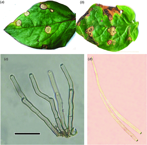
Spots on Basella alba were found on both sides of the leaves. Spots were circular to subcircular, 1–10 mm wide, at first uniformly reddish brown, later becoming blackish brown, with grey centres and reddish purple borders up to 10 mm wide (Fig. 1a, b). Dense fascicles of conidiophores were visible as minute black dots on the leaf spots. Sporulation was amphigenous, stromata or a few brown cells were present; conidiophores 1–10 in a fascicle, pale olevaceous brown, uniform in colour and width, not branched, straight or mildly geniculate with thickened conidial scars, sparingly septate, 50–140 × 3–9 µm (Fig. 1c). Conidia were hyaline, acicular, straight to slightly curved, indistinctly multiseptate, acute at the apex, truncate at the base with a thickened hilum, 45–120 × 3–8 µm (Fig. 1d). On the basis of these observations the fungus was identified as C. basellae-albae based on the description given by Srivastava et al. (1994).

|
Crous and Braun (2003) stated that C. basellae-albae was close or identical to C. apii s. lat. except that C. apii s. lat. has longer conidia (48–340 µm) and narrower conidiophores (1.5–2 µm) than C. basellae-albae. Disease specimens of C. basellae-albae have been deposited in the Mycological Herbarium, University of the Philippines Los Baños with reference collection accession number CALP 11735 on B. alba and CALP 11674 on B. rubra. This is the first record of C. basellae-albae from the Philippines.
References
Crous PW, Braun U (2003) ‘Mycosphaerella and its anamorphs: 1. Names published in Cercospora and Passalora CBS Biodiversity Series 1.’ (Centraalbureau voor Schimmel cultures: Utrecht) 569pp.ECHO (2006) ‘Malabar Spinach ECHO plant information sheet.’ (ECHO: North Fort Myers, FL). Available at http://www.echonet.org [Verified 10 September 2010].
Meeboon J, Hidayat I, To-anun C (2007) Cercosporoid fungi from Thailand 3. Two new species of Passalora and six new records of Cercospora. Mycotaxon 102, 139–145.
Srivastava RK, Narayan S, Srivastava AK (1994) New species of Cercospora from North-eastern Uttar Pradesh. Indian Phytopathology 47, 226–231.


