

Alternaria leaf spot, caused by Alternaria thunbergiae, recorded for the first time on Thunbergia alata from Brazil
M. P. Melo A , D. J. Soares B C , J. S. P. Araújo A and G. O. Tostes AA Departamento de Fitotecnia, Instituto de Agronomia, Universidade Federal Rural do Rio de Janeiro, 23890-000, Seropédica, RJ, Brazil.
B Embrapa Algodão, Rua Osvaldo Cruz, 1143, Centenário, 58428-095, Campina Grande, PB, Brazil.
C Corresponding author. Email: dartjs@yahoo.com.br
Australasian Plant Disease Notes 4(1) 23-25 https://doi.org/10.1071/DN09010
Submitted: 18 February 2009 Accepted: 6 March 2009 Published: 23 March 2009
Abstract
Alternaria thunbergiae is recorded as the cause of leaf spots on Thunbergia alata (Acanthaceae), for the first time from Brazil.
Thunbergia alata Bojer ex Sims, popularly known as black-eyed Susan vine, is native to tropical Africa and grown worldwide as an ornamental. It is also considered a potential weed in Brazil, where it invades orchards and perennial crops mainly in coastal regions (Lorenzi 2000), and in Australia (Batianoff and Butler 2002). According to Starr et al. (2003) in Hawaii T. alata is a noxious weed and ‘has an aggressive habit, climbs on other vegetation and forms a dense blanket over large areas’.
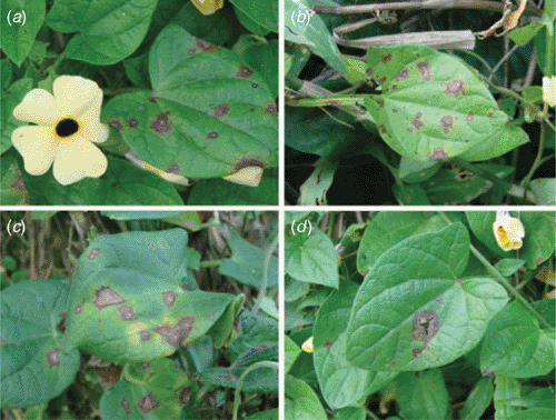
In August 2008, in the campus of the Universidade Federal Rural do Rio de Janeiro, plants of T. alata were found showing irregular, elliptic or circular leaf spots, up to 1.0 cm diameter, dark-grey to brown, with a darker border followed by a indistinct margin, mostly with a small white centre, sometimes causing the rupture of the foliar blade, rarely coalescing (Fig. 1).
|
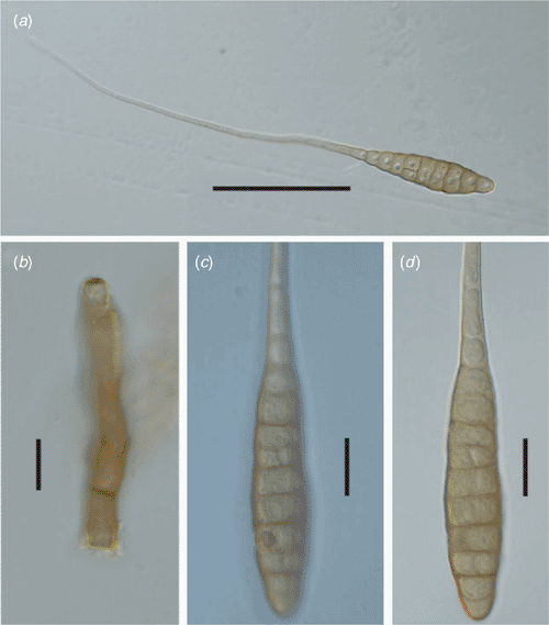
When infected tissue was examined under a stereomicroscope, typical Altenaria conidia were consistently observed on the lesions. The fungus was isolated, with the help of a fine needle, by direct transfer of conidia to plates with potato dextrose agar (PDA). Fungal structures (conidia and conidiophore) were scraped and transferred to microscope slides containing a drop of lactic acid for observation under a compound microscope. The fungus showed the following morphology: conidiophores subcylindric, straight, mostly geniculate, 50–100 × 5–8 µm, 1–5 septate, conidial scars terminal to intercalary, percurrent proliferation, pale brown to brown, smooth; conidia solitary, rarely in short chains with two conidia, with 8–10 transverse and 1–4 longitudinal septa, body 90–110 × 21–27 µm, beak up to 250 µm long, pale brown to brown, walls somewhat verruculose (Fig. 2).
|
A sample of diseased material was collected, dried in a plant press, and deposited at the herbarium of the Universidade Federal de Viçosa (Herbarium VIC. 30700). To prove pathogenicity the fungus was cultivated on plates with PDA maintained in a growth chamber at 27°C and 12 h of light. A conidial suspension with 104 spores/mL, prepared from 10-day-old colonies, was brush-inoculated on both sides of 20 healthy leaves of plants about 2 months old. After inoculation the plants were maintained for 72 h in a dew chamber, and later transferred to a greenhouse maintained at 25 ± 2°C. After 20 days the fungus was reisolated from similar symptoms to those initially observed in the field. Inoculation with mycelial discs, from cultures as described above, produced disease symptoms 12 days after inoculation on wounded leaves and 15 days on non-wounded leaves. The wounds consisted of several perforations of the leaves with a fine needle where the mycelial discs were deposited.
Based on the host, biometric and morphological data, the fungus was identified as Alternaria thunbergiae E.G. Simmons & Alcorn. This fungus was recently described from Australia, but according to Simmons (2007) this species is also known from Florida (USA). The fungal structures were mounted in lactic acid, without dye, covered with a glass slip and gently heated as described by Simmons (2007). The Brazilian specimen has narrower conidia than the type specimen from Australia (27–32 µm) and wider conidiophores than the type (3–4 µm). This discrepancy may be because our measurements were based on fungal structures (conidia and conidiophores) obtained directly from leaf lesions. We could not reproduce the standard conditions for the examination of living isolates as proposed by Simmons (2007) who based his description of A. thunbergiae on conidia and conidiophores obtained from culture. No previous recorded of any Alternaria species on T. alata was found in Brazil or South America (Embrapa 2008; Farr et al. 2008). This is the first record of A. thunbergiae from Brazil and also from South America.
Acknowledgements
The first author thanks the Fundação Carlos Chagas Filho de Amparo à Pesquisa do Estado do Rio de Janeiro (FAPERJ) for a scholarship. The second author thanks the Conselho Nacional de Desenvolvimento Científico e Tecnológico (CNPq) for financial support.
Batianoff GN, Butler DW
(2002) Assesment of invasive naturalized plants in south-east Queensland. Appendix. Plant Protection Quarterly 17, 27–34.
(verified 25 November 2008).