Seiridium cupressi is the common cause of cypress canker in south-eastern Australia
J. H. CunningtonDepartment of Primary Industries, Knoxfield Centre, Private Bag 15, Ferntree Gully Delivery Centre, Vic. 3156, Australia. Corresponding author. Email: james.cunnington@dpi.vic.gov.au
Australasian Plant Disease Notes 2(1) 53-55 https://doi.org/10.1071/DN07014
Submitted: 27 February 2007 Accepted: 6 March 2007 Published: 2 April 2007
Abstract
Cypress canker is a common disease in south-eastern Australia. The taxonomy of the Seiridium species that cause the disease has been debated due to variable morphological characters, but in Australia most workers have followed H.J. Swart and called the pathogen Seiridium unicorne. Using β-tubulin gene intron sequences, five Australian cultures were determined to be S. cupressi, rather than S. unicorne. Morphological identification of a range of herbarium specimens confirmed that S. cupressi is the common cause of cypress canker in south-eastern Australia.
Cypress canker is a serious disease of the Cupressaceae in many parts of the world (Graniti 1998). It is caused by several species of Seiridium. The identification and taxonomy of the species involved has been confused due to variable morphological characters. While some authors suggested that there was only one species responsible (Swart 1973), others believe that three species should be recognised, S. cardinale, S. cupressi and S. unicorne, based on the morphology of the conidial appendages (Boesewinkel 1983; Graniti 1998). The conidial appendages of S. cupressi follow the curve of the conidia; those of S. unicorne are usually perpendicular to the conidium; the conidia of S. cardinale lack appendages. Confusion has existed because the shape and length of the appendages is often quite variable. Barnes et al. (2001) used β-tubulin gene and histone gene sequences to show that there are three separate species but did not include any Australian isolates. The identity of the Seiridium species that cause cypress canker in Australia has been subject to several different opinions, which are discussed below.
Swart (1973) provided the first study of the cypress canker fungi in Australia, based largely on isolates from Melbourne. He suggested that S. unicorne, S. cardinale and S. cupressi might be morphological variants of the one species, and used the name S. unicorne for Australian isolates. Pathologists in Australia have generally followed this, with most cypress canker specimens in the plant pathology herbaria DAR (DPI NSW) and VPRI (DPI Victoria) labelled as S. unicorne. Boesewinkel (1983) also examined Australian specimens, but unlike Swart, believed that there were three species of Seiridium that cause cypress canker, of which both S. cupressi and S. cardinale occur in Australia. While there are some specimens labelled S. cardinale in Australian herbaria, the name S. cupressi seems never to have been used in Australia. The teleomorph of S. cupressi is Lepteutypa cupressi. The teleomorphs of the other Seiridium species are not known. Swart (1973) reported that the Lepteutypa state of the cypress canker fungus is common in Melbourne.
The identity of the fungus in Australia became more confused when Graniti and Frisullo (1991) noted that Australian isolates, which they were calling S. cupressi, differed in nutrient requirements, conidial morphology and temperature requirements from Greek isolates of S. cupressi. They thought that the Australian fungus was an undescribed species, and informally introduced the name ‘S. swartii’. The aim of this study was to re-examine a range of Australian specimens of the cypress canker fungus, using DNA sequence data where cultures were available, to determine their identity.
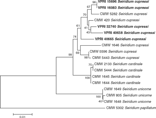
Five Australian cultures of Seiridium were obtained. Two of these were collected by H.J. Swart and were located in the culture collection of VPRI (15696 and 16083). Three recent Victorian isolates, VPRI 32740, 40658 and 40665 were obtained from fresh cypress canker samples submitted to DPI Victoria Crop Health Services in 2006. The fungus was isolated by directly removing spores from infected plant material and plating onto potato dextrose agar (PDA). DNA was extracted from the cultures using a DNeasy™ Plant Mini Kit (Qiagen) according to the manufacturer’s instructions. Two intron-containing regions of the β-tubulin gene were amplified and sequenced according to Barnes et al. (2001). The sequences have been deposited on GenBank under accession EF517786–EF517790. They were aligned with those sequences obtained by Barnes et al. (2001) and a minimum evolution tree was constructed with MEGA2 (Kumar et al. 2001) using the Kimura-2-parameter method, a complete deletion of gapped sites and 1000 bootstrap replicates (Fig. 1). The alignment was 867 bases in length. The phylogenetic tree shows all five Australian isolates clustering with the S. cupressi group with a bootstrap confidence of 74. Cultures grown on PDA in the dark were an off-white colour. Boesewinkel (1983) found that S. cupressi was off-white when grown in the dark, while S. unicorne was green when grown in the dark.

|
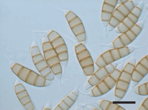
The morphology of the five cultures and a range of herbarium specimens from DAR and VPRI were examined. Material was mounted in 60% or 100% lactic acid and warmed to boiling and examined using interference contrast. The conidial appendages from all the cultures and most of the herbarium specimens were found to follow the curve of the conidia (Fig. 2) as is typical for S. cupressi. Herbarium specimens of S. cupressi from south-eastern Australia were examined on Cupressus (VPRI 21924, 31573, 32082), Sequoiadendron (VPRI 24940), Cupressocyparis (VPRI 22467), Chamaecyparis (DAR 7063, 22289, 56229), Thuja (VPRI 22447) and Platycladus (DAR 64910). Several additional specimens on Cupressus (VPRI 21172, 21764, 22475, DAR 73128) were of poor quality and conidial appendages were difficult to observe. VPRI 21639 (on Cupressus) had equal proportions of conidia with curved and perpendicular conidial appendages suggesting that it could be either S. cupressi or S. unicorne. In contrast, DAR 58443 (also on Cupressus) had short conidial appendages and appeared intermediate between S. cardinale and S. cupressi.

|
The morphological, cultural and molecular information obtained in this study demonstrates that S. cupressi is the common cause of cypress canker in south-eastern Australia. While S. unicorne and S. cardinale may be present in Australia, no specimens in this study were confidently identified as either of these species. The molecular phylogenetic analysis showed the Australian isolates of S. cupressi to be distributed amongst S. cupressi isolates from other parts of the world and does not support the suggestion that they constitute a fourth species ‘S. swartii’. The phylogenetic analysis showed that S. cupressi was more genetically variable than the other two species. This may be the reason for the morphological and biochemical differences between Australian and Greek isolates (Graniti and Frisullo 1991).
Acknowledgements
I thank Sri Kanthi de Alwis and Ramez Aldaoud for providing me with fresh material and Rodney Jones for his technical assistance.
Barnes I,
Roux J, Wingfield MJ
(2001) Characterisation of Seiridium spp. associated with cypress canker based on β-tubulin and histone sequences. Plant Disease 85, 317–321.

Boesewinkel HJ
(1983) New records of the three fungi causing cypress canker in New Zealand, Seiridium cupressi (Guba) comb. nov. and S. cardinale on Cupressocyparis and S. unicorne on Cryptomeria and Cupressus. Transactions of the British Mycological Society 80, 544–547.

Graniti A
(1998) Cypress canker: a pandemic in progress. Annual Review of Phytopathology 36, 91–114.
| Crossref | GoogleScholarGoogle Scholar | PubMed |

Graniti A, Frisullo S
(1991) Comparison of Greek and Australian isolates of Lepteutypa cupressi. Phytoparasitica 19, 261.

Kumar S,
Tamura K,
Jakobsen IB, Nei M
(2001) MEGA2: Molecular Evolutionary Genetics Analysis software. Bioinformatics 17, 1244–1245.
| Crossref | GoogleScholarGoogle Scholar | PubMed |

Swart HJ
(1973) The fungus causing cypress canker. Transactions of the British Mycological Society 61, 71–82.


