277 EFFECTS OF ERYTHROCYTE AND ERYTHROCYTE HEMOLYSATE ON BOVINE PREIMPLANTATION EMBRYO DEVELOPMENT IN VITRO UNDER REACTIVE OXYGEN SPECIES CONDITION
A. Ideta A , K. Tsuchiya A , Y. Nakamura A , M. Urakawa A , M. Murakami A , K. Hayama A and Y. Aoyagi AZen-noh Embryo Transfer Center, Kamishihoro, Hokkaido, Japan
Reproduction, Fertility and Development 22(1) 295-296 https://doi.org/10.1071/RDv22n1Ab277
Published: 8 December 2009
Abstract
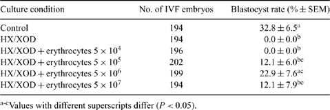
Reactive oxygen species (ROS) damage preimplantation embryos by increasing DNA fragmentation, leading to early embryonic death. Erythrocytes have been shown to protect other cells and tissues against ROS. In mice, erythrocytes were recently found to improve the early development of embryos by their antioxidant effect. The purpose of the present study was to examine the effect of erythrocytes on the in vitro development of bovine IVF embryos in medium supplemented with ROS. COCs were aspirated from ovaries collected from a local slaughterhouse and were cultured for 22 h in TCM-199 containing 5% fetal bovine serum. IVF was performed using an IVF100 (Research Institute for the Functional Peptides, Yamagata, Japan) according to the manufacturer’s instructions. In experiment 1, IVF embryos were cultured in CR1aa medium supplemented with an oxidizing agent, 0.5 mM hypoxanthine and 0.01 U mL-1 xanthine oxidase (HX/XOD), in the presence and absence of erythrocytes (5 × 10⁴, 5× 10⁵, 5×10⁶, and 5 × 10⁷ erythrocytes mL-1). In experiments 2 and 3, the development of embryos under the condition without ROS was assessed in the presence and absence of erythrocytes (5 × 10⁶ erythrocytes mL-1) or erythrocyte hemolysate (hemoglobin concentration of 1.9 g L-1), respectively. At 7 days after in vitro culture, the development to the blastocyst stage of IVF embryos was examined using a stereomicroscope. Data were analyzed using Fisher’s PLSD test and Student’s t-test In experiment 1, the presence of HX/XOD significantly inhibited embryo development to the blastocyst stage in vitro (P < 0.05). The addition of erythrocytes to medium supplemented with HX/XOD markedly improved preimplantation development (Table 1). In experiments 2 and 3, supplementation of erythrocytes or erythrocyte hemolysate promoted the development of embryos to the blastocyst stage (experiment 2: erythrocyte 42.4 ± 3.1%, control 28.5 ± 5.7%, P < 0.1; experiment 3: erythrocyte hemolysate 39.1 ± 3.3%, control 30.2 ± 1.0%, P < 0.1). In conclusion, we suggest that the addition of erythrocytes to culture medium can counteract the negative effects of ROS on embryo development and blastocyst formation.

|


