74 EFFECT OF β-MERCAPTOETHANOL ON THE VITRIFICATION CRYOTOLERANCE OF BOVINE IN VITRO-PRODUCED EMBRYOS
E. S. Ribeiro A , M. C. Gonçalves A , M. C. Pedrotti A , L. T. Martins A , R. P. C. Gerger A B , F. K. Vieira A , K. C. S. Tavares A , M. Bertolini A and A. Mezzalira AA Center of Agroveterinarian Sciences, Santa Catarina State University, Lages, SC, Brazil;
B FMVZ, University of São Paulo, São Paulo, SP, Brazil
Reproduction, Fertility and Development 21(1) 137-138 https://doi.org/10.1071/RDv21n1Ab74
Published: 9 December 2008
Abstract
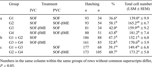
The control of oxidative processes in in vitro production (IVP) systems by the use of additives may be an alternative approach to improve embryo cryotolerance. The aim of this study was to verify the effect of β-mercaptoethanol (βME) on the cryotolerance of bovine IVP embryos. In 7 replications, and following IVM-IVF, presumptive zygotes (n = 3735) were in vitro-cultured in SOF medium supplemented or not with 100 μm βME (IVC treatment), at 38.5°C and high humidity. The initial 24 h of IVC was performed in 5% CO2 in air, with the remaining 6 days of IVC carried out in 5% CO2, 5% O2, and 90% N2. On Day 7, resulting blastocysts and expanded blastocysts were vitrified in glass micropipettes in a solution with 20% ethylene glycol + 20% propylene glycol. After warming, embryos were randomly allocated to 1 of 2 sub-groups for an additional 72 h of IVC to the hatching blastocyst (HBL) stage, in fresh SOF medium supplemented or not with 100 μm βME (PVC treatment), at 38.5°C, high humidity and 5% CO2. Experimental groups were as follows: G1 (βME-free medium during IVC and PVC); G2 (βME only during PVC); G3 (βME only during IVC); and G4 (βME during IVC and PVC). Cleavage (Day 2) and blastocyst (Day 7) rates in the IVC treatment and hatching rates (Days 7 to 9) for the PVC treatment were analyzed by the chi-square test, for P < 0.05. Total cell number (TCN) estimated by fluorescence staining in HBL derived from vitrified and nonvitrified embryos was analyzed by ANOVA. The use of βME during IVC did not affect cleavage rates (βME-free, 1491/1858, 80.2% v. βME, 1522/1877, 81.1%), but negatively affected development to the blastocyst stage (βME-free, 813/1858, 43.8% v. βME, 525/1877, 28.0%). Following vitrification, however, βME supplementation during PVC improved hatching rates (G2, 58.1% and G4, 63.8%) compared with groups without the additive (G1, 36.6% and G3, 42.0%). In addition, the presence of βME either during IVC or PVC, or during both culture periods, increased TCN in HBL from vitrified embryos (Table 1). The use of βME during IVC, irrespective of the presence of βME during the PCV period, caused an increase in TCN in HBL in G3 + G4, with no effects on hatching rates (Table 1b), whereas the addition of βME during PVC, irrespective of the presence of βME during the IVC period, resulted in greater hatching rates and TCN in HBL in G2 + G4 than in G1 + G3 (Table 1). In conclusion, the addition of βME during the IVC period did not affect cleavage, but reduced blastocyst yield. Despite that, βME supplementation during the IVC period appeared to have increased the cryotolerance of the resulting blastocysts, expressed by greater TCN in HBL, whereas βME supplementation during the PVC period also improved embryo survival to the vitrification process, manifested by greater hatching rates and TCN in HBL.

|
This study was supported by a grant from CNPq/Brazil.


