First report of Colletotrichum phormii the cause of anthracnose on Phormium tenax in Australia
H. Golzar A B and C. Wang AA Department of Agriculture and Food Western Australia, Bentley Delivery Centre, WA 6983, Australia.
B Corresponding author. Email: hgolzar@agric.wa.gov.au
Australasian Plant Disease Notes 5(1) 110-112 https://doi.org/10.1071/DN10040
Submitted: 12 July 2010 Accepted: 22 September 2010 Published: 4 October 2010
Abstract
In February 2010, anthracnose symptoms were observed on Phormium tenax in Perth, WA, Australia. Based on disease symptoms, the morphological characteristics of the isolated fungus, pathogenicity tests and sequencing of the internal transcribed spacer region of the rDNA, Colletotrichum phormii was identified. This is the first report of C. phormii causing anthracnose on Phormium tenax in Australia.
Phormium species belonging to the Agavaceae family have been widely distributed in the temperate regions of the world. Phormium tenax (New Zealand flax) is a perennial evergreen and is native to New Zealand (Wehi and Clarkson 2007). Colletotrichum species cause the disease commonly known as anthracnose with significant economic losses relating to a wide range of plant species worldwide. Colletotrichum gloeosporioides (teleomorph Glomerella cingulata) and C. acutatum (teleomorph Glomerella acutata) are the most common species causing anthracnose on various plant species (Mordue 1971; Dyko and Mordue 1979; Bailey and Jeger 1992). Colletotrichum species have been reported on specific genera in the Agavaceae including C. agaves on Agave, C. dracaenophilum on Dracaena, and C. phormii on Phormium. C. phormii has been known to be the slowest growing and longest-spored species of Colletotrichum on Agavaceae and commonly attacks Phormium species, particularly P. tenax worldwide (Farr et al. 2006; Hyde et al. 2009). A Colletotrichum sp. with similar conidia has been reported on Dracaena sanderiana from China (Farr et al. 2006).
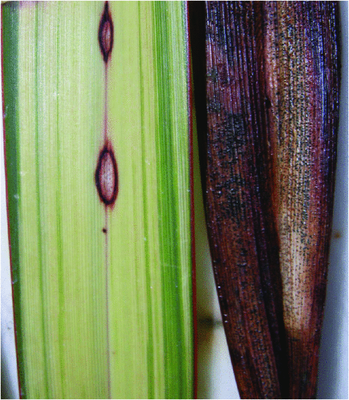
Anthracnose symptoms were observed on leaves of P. tenax in Perth, WA, Australia. Typical symptoms appeared as dark brown lesions with discoloration of central tissues that eventually coalesced to form large irregular necrotic areas. In older lesions black sub-epidermal acervuli formed, with erumpent through the epidermis with no obvious setae (Fig. 1). Fifty sections of plant tissue surrounding lesions were surface-sterilised by immersion in a 1.25% aqueous solution of sodium hypochlorite for 1 min, rinsed in sterile water and dried in a laminar flow cabinet. The sterilised leaf pieces were then either placed on potato dextrose agar (PDA) and incubated at 22 ± 3°C, with developing fungal colonies sub-cultured onto PDA and then single-spored to obtain pure cultures; or placed in trays on moist filter paper and incubated at 25°C with a 12-h dark and light cycle. Colletotrichum sp. was consistently isolated and the growth rate, colony morphology and morphological characteristics of the isolated fungus were determined for 10 isolates.

|
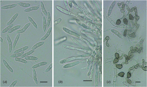
Colonies were pale brown turning olivaceous-grey after 2 weeks and developed slowly on PDA with an average diameter of 28 mm after 7 days. Acervuli were black, 250–400 µm without setae on plant material and in culture. Conidiogenous cells were cylindrical, 12–24 × 3.5–5 µm. The conidia were hyaline, cylindrical to fusiform, slightly curved with truncate base and rounded apex and measured 17.5–30.5 × 4–7.5 µm. Appressoria were terminal or lateral, obpyriform to irregularly lobed, 6–11 × 5–9 µm (Fig. 2). Both cultural and morphological characteristics of the isolates were similar to those described for C. phormii (Farr et al. 2006).The teleomorph, Glomerella phormii, was not observed.

|
Two representative isolates of morphologically identified Colletotrichum phormii were grown on PDA for 10 days at 21°C. Genomic DNA was extracted from fungal mycelium with a DNeasy Plant Mini Kit (Qiagen, Melbourne, Vic., Australia) according to the manufacturer’s instructions. Amplification of the internal transcribed spacer (ITS)1 and ITS2 regions flanking the 5.8S rRNA gene was carried out with universal primers ITS1 and ITS4 according to the published protocol (White et al. 1990). The polymerase chain reaction (PCR) product was purified and sequenced on an ABI 3730 DNA Sequencer (Applied Biosystems, Melbourne, Vic., Australia) at Murdoch University, Perth, WA, Australia. The sequences of the ITS1 and ITS2 regions were identical to the sequences from the same region of C. phormii in the NCBI database (www.ncbi.nlm.nih.gov/) as published by Farr et al. (2006).
Pathogenicity tests were performed on 24 P. tenax seedlings using two representative isolates of C. phormii. Plants were inoculated (leaves and stems) by either wounding or non-wounding. Wounding was used to accelerate symptom development as C. phormii has been reported to be the slowest growing species of Colletotrichum on Agavaceae family (Farr et al. 2006). A conidial suspension (106 spores/mL) was prepared in sterile water using a 10-day-old culture of each isolate grown on PDA. For the wounding inoculation test (8 plants), the conidial suspension was dropped on the leaf and stem sites (5 µL per site/10 sites per plant) after pin-prink wounding using a syringe needle. For the non-wounding inoculation test (8 plants) the conidial suspension was sprayed on the plant surface to runoff. Controls (8 plants) were inoculated using sterilised water as described previously (Lubbe et al. 2006). All plants were placed under mist for 48 h and then moved to a growth room chamber at 22 ± 1°C. Disease symptoms were observed 2 weeks post-inoculation and symptom development was accelerated on wound-inoculated compared with non-wound-inoculated plants. Control plants remained asymptomatic.
Koch’s postulates were fulfilled by reisolation of C. phormii. A culture of C. phormii, the morphological identity of which was confirmed via ITS sequencing, was deposited in the Western Australia Plant Pathogen Collection (WAC 12416). To our knowledge, this is the first report of C. phormii causing anthracnose on P. tenax in Australia.
Acknowledgement
The authors would like to thank Ms Paula Mather for technical assistance.
References
Bailey JA, Jeger MJ (1992) ‘Colletotrichum: Biology, Pathology and Control.’ (CAB International: Wallingford UK)Dyko BJ, Mordue JEM (1979) Colletotrichum acutatum. CMI (Commonwealth Mycological Institute) Description of Pathogenic Fungi and Bacteria. No, 630.
Farr DF, Aime MC, Rossman AY, Palm ME (2006) Species of Colletotrichum on Agavaceae. Mycological Research 110, 1395–1408.
| Species of Colletotrichum on Agavaceae.Crossref | GoogleScholarGoogle Scholar | 1:CAS:528:DC%2BD2sXhsVOls74%3D&md5=bd63929746f7002e5c5b97bb13f17daaCAS | 17137776PubMed |
Hyde KD, Cai L, Cannon PF, Crouch JA, Crous PW, Damm U, Goodwin PH, Chen H, Johnston PR, Jones EBG, Liu ZY, McKenzie EHC, Moriwaki J, Noireung P, Pennycook SR, Pfenning LH, Prihastuti H, Sato T, Shivas RG, Tan YP, Taylor PWJ, Weir BS, Yang YL, Zhang JZ (2009) Colletotrichum – names in current use. Fungal Diversity 39, 147–182. Available online at www.fungaldiversity.org/fdp/sfdp/FD39-7.pdf
Lubbe CM, Denman S, Lamprecht SC, Crous PW (2006) Pathogenicity of Colletotrichum species to Protea cultivars. Australasian Plant Pathology 35, 37–41.
| Pathogenicity of Colletotrichum species to Protea cultivars.Crossref | GoogleScholarGoogle Scholar |
Mordue JEM (1971) Glomerella cingulata. CMI (Commonwealth Mycological Institute) Description of Pathogenic Fungi and Bacteria. No, 315.
Wehi PM, Clarkson BD (2007) Biological flora of New Zealand. 10. Phormium tenax, harakeke, New Zealand flax. New Zealand Journal of Botany 45, 521–544.
| Biological flora of New Zealand. 10. Phormium tenax, harakeke, New Zealand flax.Crossref | GoogleScholarGoogle Scholar |
White TJ, Bruns T, Lee S, Taylor J (1990) Amplification and direct sequencing of fungal ribosomal RNA genes for phylogenetics. In ‘PCR protocols: a guide to methods and applications’. (Eds MA Innis, DH Gelfand, JJ Sninsky, TJ White) pp. 315–322. (Academic Press: San Diego)


